Japanese
English
- 有料閲覧
- Abstract 文献概要
- 1ページ目 Look Inside
- 参考文献 Reference
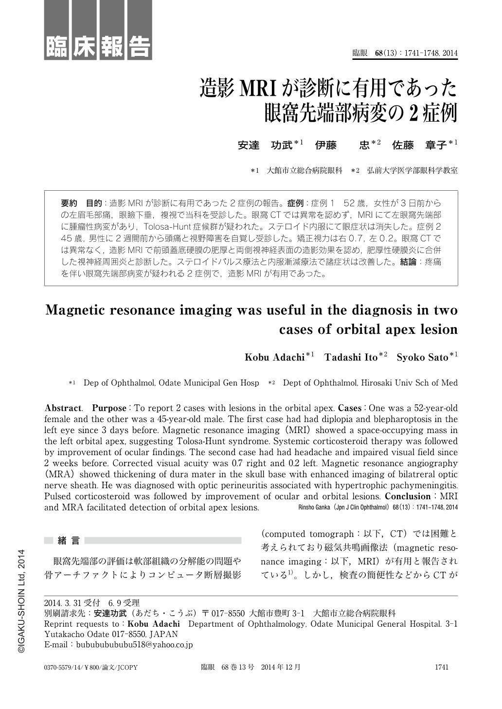
要約 目的:造影MRIが診断に有用であった2症例の報告。症例:症例1 52歳,女性が3日前からの左眉毛部痛,眼瞼下垂,複視で当科を受診した。眼窩CTでは異常を認めず,MRIにて左眼窩先端部に腫瘤性病変があり,Tolosa-Hunt症候群が疑われた。ステロイド内服にて眼症状は消失した。症例2 45歳,男性に2週間前から頭痛と視野障害を自覚し受診した。矯正視力は右0.7,左0.2。眼窩CTでは異常なく,造影MRIで前頭蓋底硬膜の肥厚と両側視神経表面の造影効果を認め,肥厚性硬膜炎に合併した視神経周囲炎と診断した。ステロイドパルス療法と内服漸減療法で諸症状は改善した。結論:疼痛を伴い眼窩先端部病変が疑われる2症例で,造影MRIが有用であった。
Abstract. Purpose:To report 2 cases with lesions in the orbital apex. Cases:One was a 52-year-old female and the other was a 45-year-old male. The first case had had diplopia and blepharoptosis in the left eye since 3 days before. Magnetic resonance imaging(MRI)showed a space-occupying mass in the left orbital apex, suggesting Tolosa-Hunt syndrome. Systemic corticosteroid therapy was followed by improvement of ocular findings. The second case had had headache and impaired visual field since 2 weeks before. Corrected visual acuity was 0.7 right and 0.2 left. Magnetic resonance angiography(MRA)showed thickening of dura mater in the skull base with enhanced imaging of bilatreral optic nerve sheath. He was diagnosed with optic perineuritis associated with hypertrophic pachymeningitis. Pulsed corticosteroid was followed by improvement of ocular and orbital lesions. Conclusion:MRI and MRA facilitated detection of orbital apex lesions.

Copyright © 2014, Igaku-Shoin Ltd. All rights reserved.


