Japanese
English
- 有料閲覧
- Abstract 文献概要
- 1ページ目 Look Inside
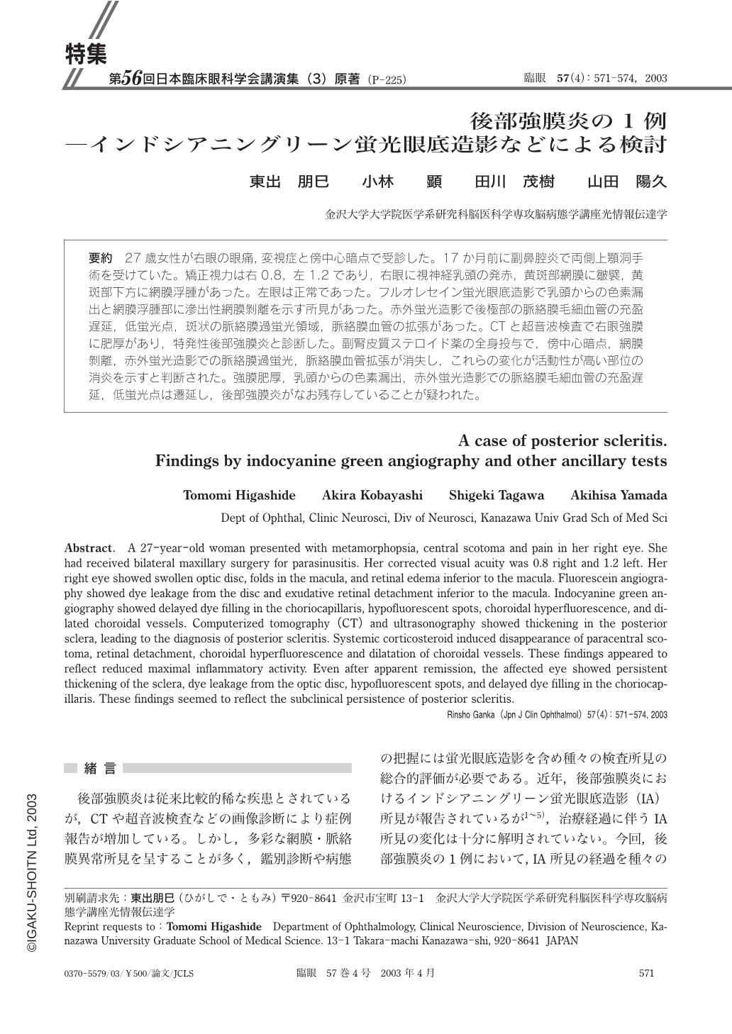
要約 27歳女性が右眼の眼痛,変視症と傍中心暗点で受診した。17か月前に副鼻腔炎で両側上顎洞手術を受けていた。矯正視力は右0.8,左1.2であり,右眼に視神経乳頭の発赤,黄斑部網膜に皺襞,黄斑部下方に網膜浮腫があった。左眼は正常であった。フルオレセイン蛍光眼底造影で乳頭からの色素漏出と網膜浮腫部に滲出性網膜剝離を示す所見があった。赤外蛍光造影で後極部の脈絡膜毛細血管の充盈遅延,低蛍光点,斑状の脈絡膜過蛍光領域,脈絡膜血管の拡張があった。CTと超音波検査で右眼強膜に肥厚があり,特発性後部強膜炎と診断した。副腎皮質ステロイド薬の全身投与で,傍中心暗点,網膜剝離,赤外蛍光造影での脈絡膜過蛍光,脈絡膜血管拡張が消失し,これらの変化が活動性が高い部位の消炎を示すと判断された。強膜肥厚,乳頭からの色素漏出,赤外蛍光造影での脈絡膜毛細血管の充盈遅延,低蛍光点は遷延し,後部強膜炎がなお残存していることが疑われた。
Abstract. A 27-year-old woman presented with metamorphopsia,central scotoma and pain in her right eye. She had received bilateral maxillary surgery for parasinusitis. Her corrected visual acuity was 0.8 right and 1.2 left. Her right eye showed swollen optic disc,folds in the macula,and retinal edema inferior to the macula. Fluorescein angiography showed dye leakage from the disc and exudative retinal detachment inferior to the macula.Indocyanine green angiography showed delayed dye filling in the choriocapillaris,hypofluorescent spots,choroidal hyperfluorescence,and dilated choroidal vessels. Computerized tomography(CT)and ultrasonography showed thickening in the posterior sclera,leading to the diagnosis of posterior scleritis. Systemic corticosteroid induced disappearance of paracentral scotoma,retinal detachment,choroidal hyperfluorescence and dilatation of choroidal vessels. These findings appeared to reflect reduced maximal inflammatory activity. Even after apparent remission,the affected eye showed persistent thickening of the sclera,dye leakage from the optic disc,hypofluorescent spots,and delayed dye filling in the choriocapillaris. These findings seemed to reflect the subclinical persistence of posterior scleritis.
Copyright © 2003, Igaku-Shoin Ltd. All rights reserved.