Japanese
English
- 有料閲覧
PPV購入案内
1,320円 (1,200円+税10%) こちらは、857ページから862ページの文献です。購入には医書.jp本会員登録が必要です。
会員登録完了後に改めて上記「購入サイトへ」を選択してください。
または「購入サイトへ」選択後に「購入ログイン」、「新規会員登録」の順に進んでください。
会員登録について詳しくはをご確認ください。
- Abstract 文献概要
- 1ページ目 Look Inside
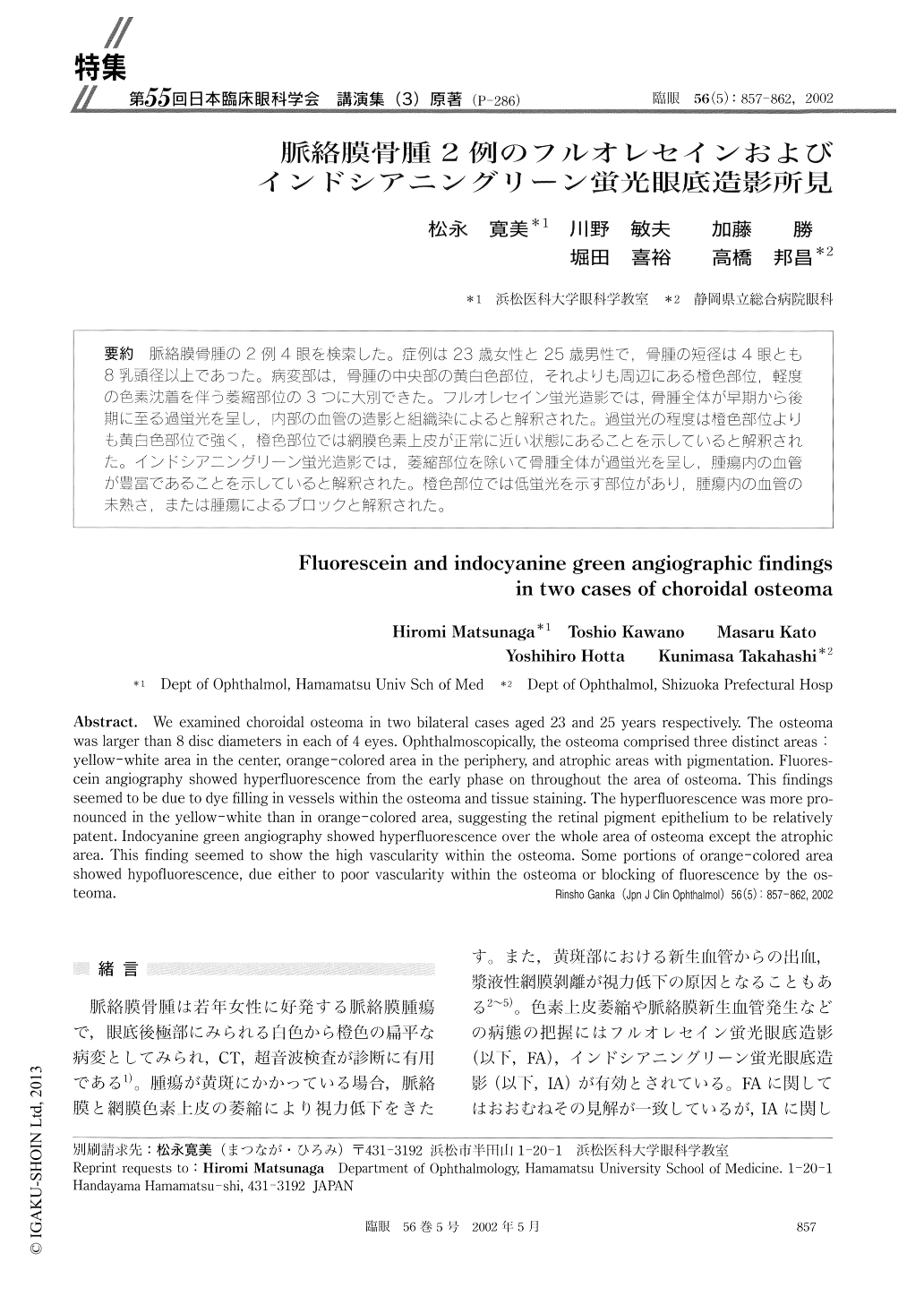
脈絡膜骨腫の2例4眼を検索した。症例は23歳女性と25歳男性で,骨腫の短径は4眼とも8乳頭径以上であった。病変部は,骨腫の中央部の黄白色部位,それよりも周辺にある燈色部位,軽度の色素沈着を伴う萎縮部位の3つに大別できた。フルオレセイン蛍光造影では,骨腫全体が早期から後期に至る過蛍光を呈し,内部の血管の造影と組織染によると解釈された。過蛍光の程度は橙色部位よりも黄白色部位で強く,橙色部位では網膜色素上皮が正常に近い状態にあることを示していると解釈された。インドシアニングリーン蛍光造影では,萎縮部位を除いて骨腫全体が過蛍光を呈し,腫瘍内の血管が豊畠であることを示していると解釈された。橙色部位では低蛍光を示す部位があり,腫瘍内の血管の未熟さ,または腫瘍によるブロックと解釈された。
We examined choroidal osteoma in two bilateral cases aged 23 and 25 years respectively. The osteoma was larger than 8 disc diameters in each of 4 eyes. Ophthalmoscopically, the osteoma comprised three distinct areas yellow-white area in the center, orange-colored area in the periphery, and atrophic areas with pigmentation. Fluores-cein angiography showed hyperfluorescence from the early phase on throughout the area of osteoma. This findings seemed to be due to dye filling in vessels within the osteoma and tissue staining. The hyperfluorescence was more pro-nounced in the yellow-white than in orange-colored area, suggesting the retinal pigment epithelium to be relatively patent. Indocyanine green angiography showed hyperfluorescence over the whole area of osteoma except the atrophic area. This finding seemed to show the high vascularity within the osteoma. Some portions of orange-colored area showed hypofluorescence, due either to poor vascularity within the osteoma or blocking of fluorescence by the os-teoma.
Copyright © 2002, Igaku-Shoin Ltd. All rights reserved.