Japanese
English
Report from overseas
米国における超音波診断(その4)—血管外科への応用
Operative ultrasonic diagnosis in the United States: Application in vascular surgery
町 淳二
1
,
Julio C. Beitler
1
,
D. Preston Flanigan
1
,
James J. Schuler
1
,
Julio C. U. Coelho
1
Junji MACHI
1
,
Bernard Sigel
1
1イリノイ大学医学部外科学教室
1Department of Surgery, University of Illinois
pp.111-118
発行日 1983年1月20日
Published Date 1983/1/20
DOI https://doi.org/10.11477/mf.1407208224
- 有料閲覧
- Abstract 文献概要
- 1ページ目 Look Inside
はじめに
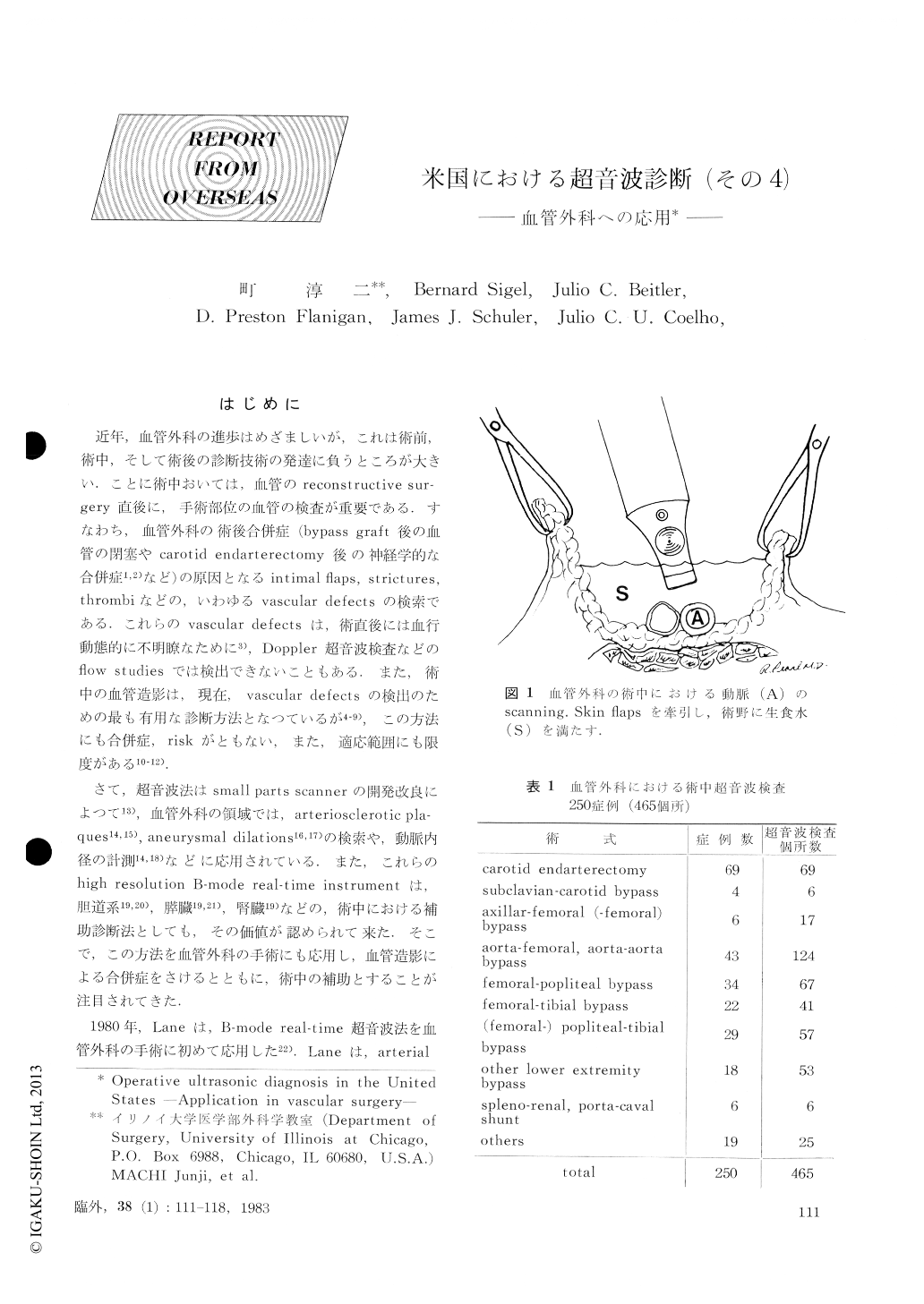
近年,血管外科の進歩はめざましいが,これは術前,術中,そして術後の診断技術の発達に負うところが大きい.ことに術中おいては,血管のreconstructive sur-gery直後に,手術部位の血管の検査が重要である.すなわち,血管外科の術後合併症(bypass graft後の血管の閉塞やcarotid endarterectomy後の神経学的な合併症1,2)など)の原因となるintimal flaps,strictures,thrombiなどの,いわゆるvascular defectsの検索である.これらのvascular defectsは,術直後には血行動態的に不明瞭なために3),Doppler超音波検査などのflow studiesでは検出できないこともある.また,術中の血管造影は,現在,vascular defectsの検出のための最も有用な診断方法となつているが4-9),この方法にも合併症,riskがともない,また,適応範囲にも限度がある10-12).
さて,超音波法はsmall parts scannerの開発改良によつて13),血管外科の領域では,arteriosclerotic plaques14,15),aneurysmal dilations16,17)の検索や,動脈内径の計測14,18)などに応用されている.

Copyright © 1983, Igaku-Shoin Ltd. All rights reserved.


