Japanese
English
- 有料閲覧
- Abstract 文献概要
- 1ページ目 Look Inside
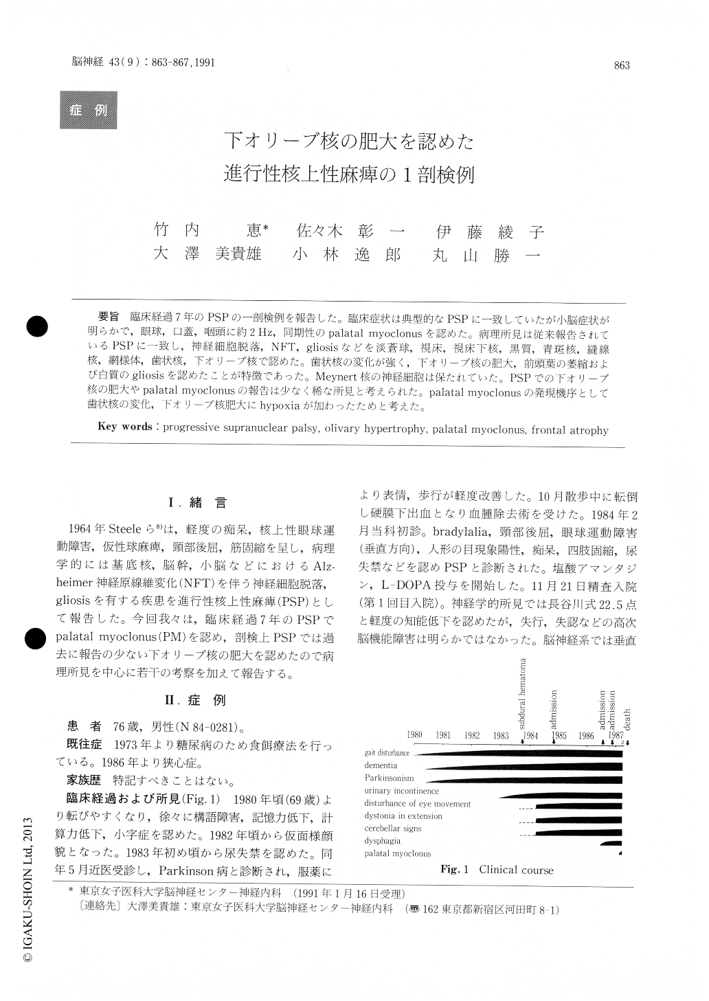
臨床経過7年のPSPの一剖検例を報告した。臨床症状は典型的なPSPに一致していたが小脳症状が明らかで,眼球,口蓋,咽頭に約2Hz,同期性のpalatal myoclonusを認めた。病理所見は従来報告されているPSPに一致し,神経細胞脱落,NFT, gliosisなどを淡蒼球,視床,視床下核,黒質,青斑核,縫線核,網様体,歯状核,下オリーブ核で認めた。歯状核の変化が強く,下オリーブ核の肥大,前頭葉の萎縮および白質のgliosisを認めたことが特徴であった。Meyllert核の神経細胞は保たれていた。PSPでの下オリーブ核の肥大やpalatal myoclonusの報告は少なく稀な所見と考えられた。palatal myoclonusの発現機序として歯状核の変化,下オリーブ核肥大にhypoxiaが加わったためと考えた。
Clinical and pathologic findings of an autopsy case of progressive supranuclear palsy (PSP) with a 7 year clinical course are described. The patient exhibited clinical findings of typical PSP, cerebellar signs and rhythmical myoclonus that was about 2 Hz and synchronous in the eyes, palate, and phar-ynx, which is so called palatal myoclonus. Patholo-gical findings compatible with those in PSP i. e. loss of nerve cells, neurofibrillary tangles (NFT) , and gliosis were found in the globus pallidum, thalamus, subthalamic nucleus, substantia nigra, locus coer-uleus, nucleus of Raphe, reticular formation, dentate nucleus, and inferior olives. Nerve cells in thenucleus basalis were preserved. Distinctive findings included marked degeneration of the dentate nucleus, prominent hypertrophy of the inferior olives, and atrophy and subcortical gliosis of the frontal lobe.
Hypertrophy of the inferior olives and palatal myoclonus represent an unusual PSP. It is presumedhypoxic injury unmasked the palatal myoclonus in this setting of dentate nucleus and inferior olivary complex degeneration.

Copyright © 1991, Igaku-Shoin Ltd. All rights reserved.


