Japanese
English
- 有料閲覧
PPV購入案内
1,320円 (1,200円+税10%) こちらは、1073ページから1079ページの文献です。購入には医書.jp本会員登録が必要です。
会員登録完了後に改めて上記「購入サイトへ」を選択してください。
または「購入サイトへ」選択後に「購入ログイン」、「新規会員登録」の順に進んでください。
会員登録について詳しくはをご確認ください。
- Abstract 文献概要
- 1ページ目 Look Inside
- 参考文献 Reference
Ⅰ.はじめに
可逆性脳血管攣縮症候群(reversible cerebral vasoconstriction syndrome:RCVS)に合併する皮質性くも膜下出血(cortical subarachnoid hemorrhage:cSAH)は発症早期に約1/3の症例で認められるが,その出血機序については明らかではない3,7).今回われわれは,雷鳴様頭痛(thunderclap headache:TH)発症後2日目の画像検査でcSAHを認め,血腫が最も厚い脳溝部に近接して急性期微小皮質梗塞を合併したと考えられるRCVSの1例を経験した.稀な症例であり,RCVSに伴うcSAHの臨床像やその発生機序について,文献的考察を加えて報告する.
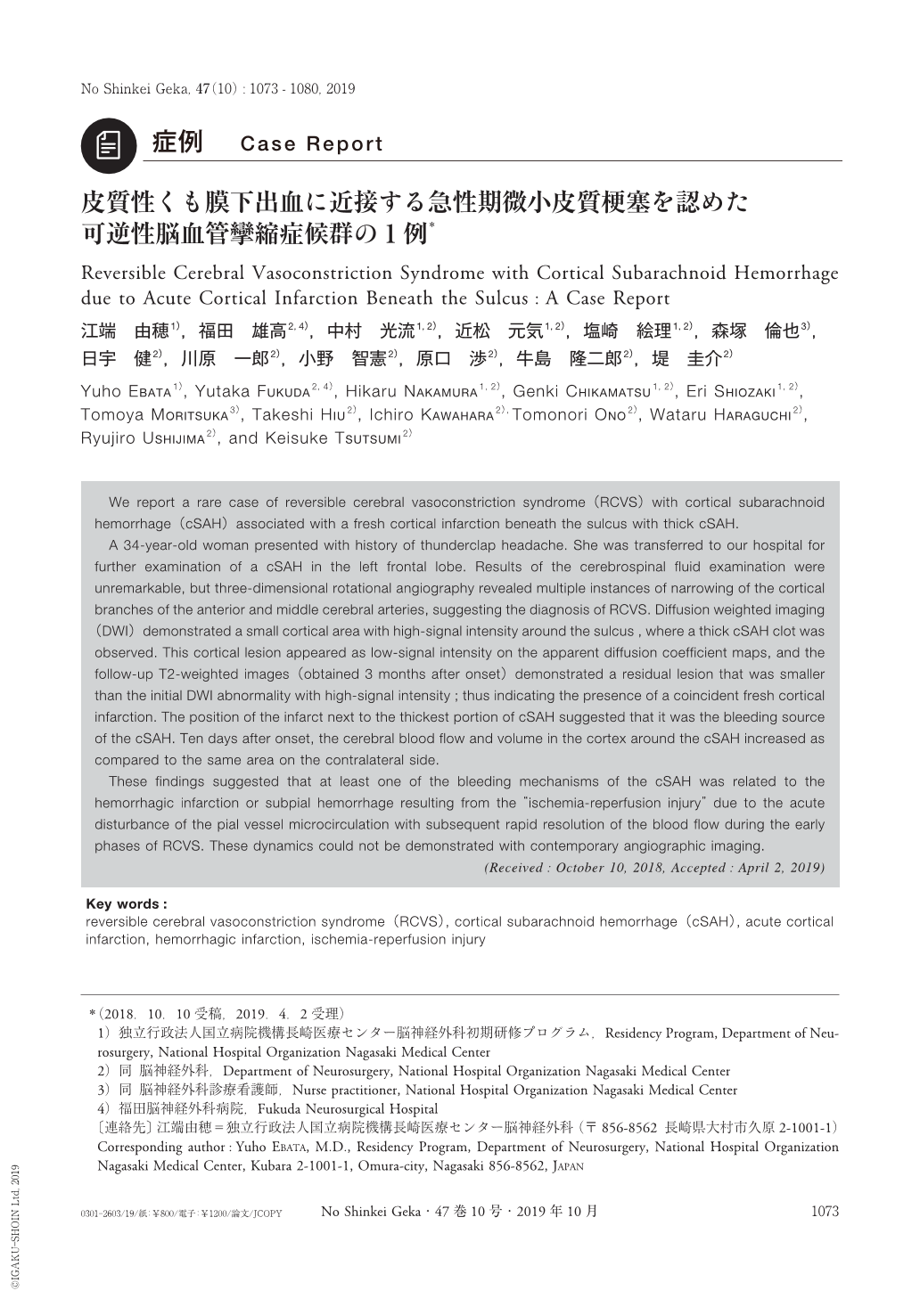
We report a rare case of reversible cerebral vasoconstriction syndrome(RCVS)with cortical subarachnoid hemorrhage(cSAH)associated with a fresh cortical infarction beneath the sulcus with thick cSAH.
A 34-year-old woman presented with history of thunderclap headache. She was transferred to our hospital for further examination of a cSAH in the left frontal lobe. Results of the cerebrospinal fluid examination were unremarkable, but three-dimensional rotational angiography revealed multiple instances of narrowing of the cortical branches of the anterior and middle cerebral arteries, suggesting the diagnosis of RCVS. Diffusion weighted imaging(DWI)demonstrated a small cortical area with high-signal intensity around the sulcus , where a thick cSAH clot was observed. This cortical lesion appeared as low-signal intensity on the apparent diffusion coefficient maps, and the follow-up T2-weighted images(obtained 3 months after onset)demonstrated a residual lesion that was smaller than the initial DWI abnormality with high-signal intensity;thus indicating the presence of a coincident fresh cortical infarction. The position of the infarct next to the thickest portion of cSAH suggested that it was the bleeding source of the cSAH. Ten days after onset, the cerebral blood flow and volume in the cortex around the cSAH increased as compared to the same area on the contralateral side.
These findings suggested that at least one of the bleeding mechanisms of the cSAH was related to the hemorrhagic infarction or subpial hemorrhage resulting from the “ischemia-reperfusion injury” due to the acute disturbance of the pial vessel microcirculation with subsequent rapid resolution of the blood flow during the early phases of RCVS. These dynamics could not be demonstrated with contemporary angiographic imaging.
Copyright © 2019, Igaku-Shoin Ltd. All rights reserved.