Japanese
English
- 有料閲覧
- Abstract 文献概要
- 1ページ目 Look Inside
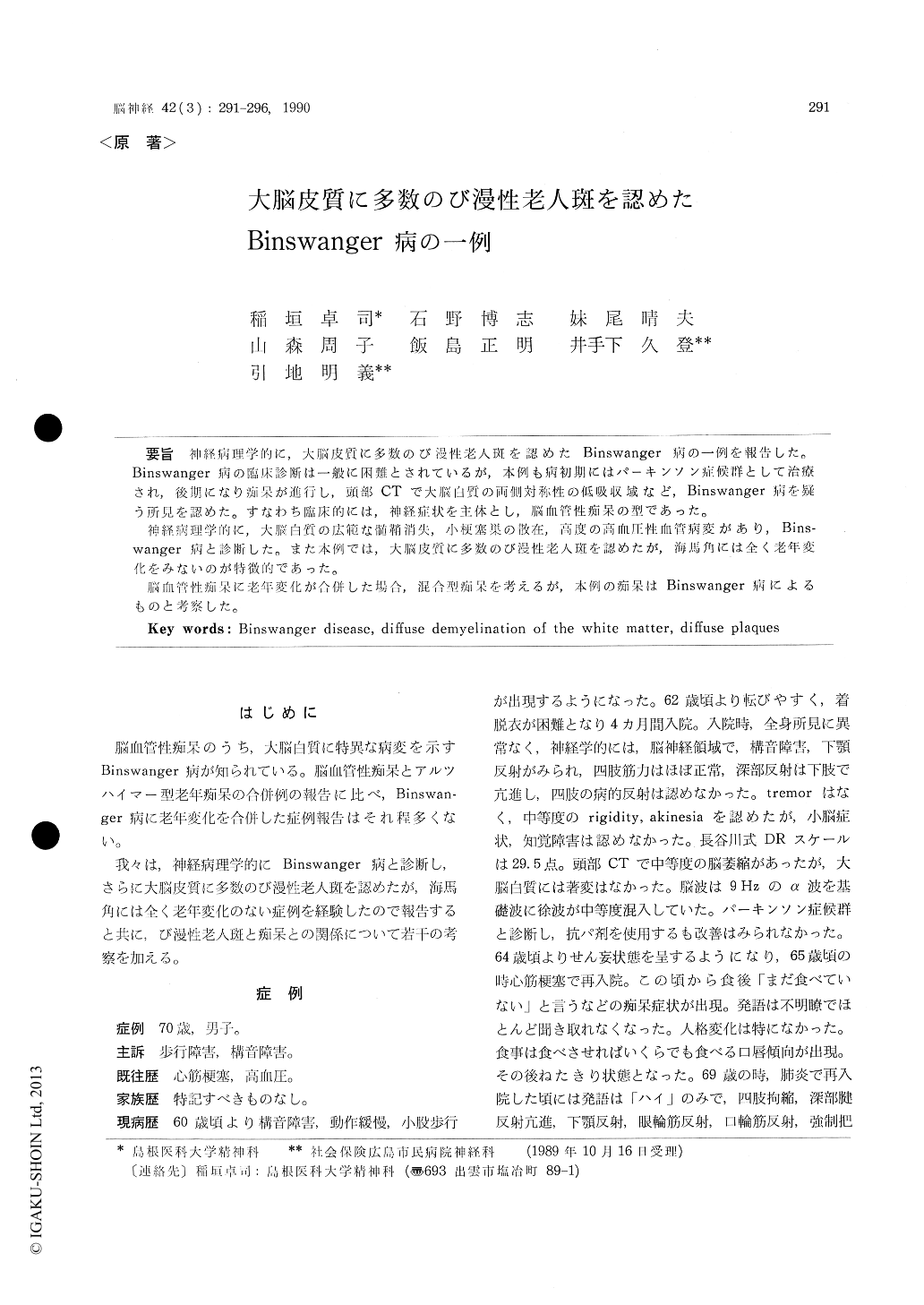
神経病理学的に,大脳皮質に多数のび漫性老人斑を露忍めたBinswanger病の一例を報告した。Binswanger病の臨床診断は一般に困難とされているが,本例も病初期にはパーキンソン症候群として治療され,後期になり痴呆が進行し,頭部CTで大脳白質の両側対称性の低吸収域など,Binswanger病を疑う所見を認めた。すなわち臨床的には,神経症状を主体とし,脳血管性痴呆の型であった。
神経病理学的に,大脳白質の広範な髄鞘消失,小梗塞巣の散在,高度の高血圧性血管病変があり,Bins—wanger病と診断した。また本例では,大脳皮質に多数のび漫性老人斑を認めたが,海馬角には全く老年変化をみないのが特徴的であった。
脳血管性痴呆に老年変化が合併した場合,混合型痴呆を考えるが,本例の痴呆はBinswanger病によるものと考察した。
A case of Binswanger disease with numerous diffuse plaques in the neocortex was reported.
This male patient had a previous history of hypertension and myocardial infarction. From the age of 60, he developed dysarthria, brady-kinesia, marche a petit pas and falling down. Neurological examination at his first admission disclosed muscular rigidity and increased jaw and deep tendon reflexes, but dementia was not found. Brain CT showed moderate brain atrophy and EEG consisted of slow wave dysrythmia. He was diagnosed of Parkinsonism and treatment started without effects.
During his second admission for the treatment of myocardial infarction, at the age of 64, deli-rium developed. Progressive dementia began and finally he was confined to bed. From the age of 69, spontaneous speech became almost lost. Contracture of the extremities, increased deep tendon reflexes and forced grasping were noted.Brain CT showed symmetrical low attenuation in the frontal and parietal white matter with moderate dilatation of the lateral ventricles.
At the age of 70, he died of general prostra-tion about ten years after the initial symptoms. Neuropathological findings :
Macroscopic findings :
The brain weighed 1300 g. Atherosclerotic changes of the large arteries at the base of the brain were moderate. Coronal sections of the brain showed moderate enlargement of the lateral ventricles with multiple small lacunes in the basal ganglia.
Microscopic findings :
Bilateral diffuse demyelination of the white matter with sparing of the U-fibers was noted. Holzer stain revealed fibrillary gliosis in the left parietal and occipital white matter. Marked adventitial fibrosis of the deep white matter arteries and terminal stages of hyalinosis of the perforating arteries were found. Basal ganglia showed status lacunaris. Bilateral pyramidal tracts were atrophic secondally.
These pathological findings were consistent with the criteria of Binswnger disease.
Characteristic findings in this case were as fol-lows.
1) There were numerous senile plaques in the cerebral neocortex without neurofibrially tangles. These plaques were stained by Hirano stain and by methenamin-Bodian stain and did not contain swollen degenerative neurites. In contrast, Bodian preparations did not demonstrate these plaques. These plaques were consistent with the so-called diffuse plaques (very primitive plaques and an initial stage of senile plaques).
2) Hippocampus was entirely free of senile degenerative changes.
We have called attention to the association of Binswanger disease and diffuse plaques. We dis-cussed that the dementia of this patient was more related to Binswanger disease process than to numerous diffuse plaques in the neocortex, as the role of diffuse plaques in dementia symptoms has not been well established.

Copyright © 1990, Igaku-Shoin Ltd. All rights reserved.


