Japanese
English
- 有料閲覧
- Abstract 文献概要
- 1ページ目 Look Inside
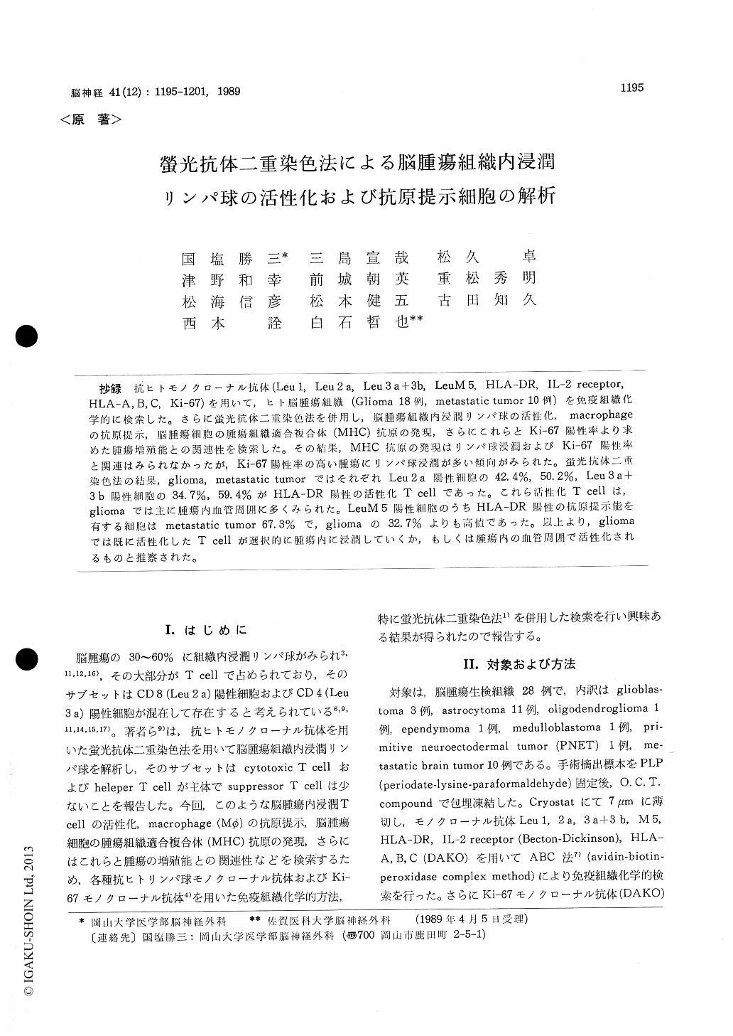
抄録 抗ヒトモノクローナル抗体(Leu 1, Leu 2a, Leu 3a+3b, LeuM 5, HLA-DR, IL−2 receptor, HLA-A, B, C, Ki−67)を用いて,ヒト脳腫瘍組織(Glioma 18例, metastatic tumor 10例)を免疫組織化学的に検索した。さらに蛍光抗体二重染色法を併用し,脳腫瘍組織内浸潤リンパ球の活性化,macrophageの抗原提示,脳腫瘍細胞の腫瘍組織適合複合体(MHC)抗原の発現,さらにこれらとKi−67陽性率より求めた腫瘍増殖能との関連性を検索した。その結果,MHC抗原の発現はリンパ球浸潤およびKi−67陽性率と関連はみられなかったが,Ki−67陽性率の高い腫瘍にリンパ球浸潤が多い傾向がみられた。蛍光抗体二重染色法の結果,glioma ,metastatic tumorではそれぞれLeu 2a陽性細胞の42.4%, 50.2%, Leu3a+3b陽性細胞の34.7%, 59.4%がHLA-DR陽性の活性化T cellであった。これら活性化T cellは,gliomaでは主に腫瘍内血管周囲に多くみられた。LeuM 5陽性細胞のうちHLA-DR陽性の抗原提示能を有する細胞はmetastatic tumor 67.3%で,gliomaの32.7%よりも高値であった。以上よりg1iomaでは既に活性化したT cellが選択的に腫瘍内に浸潤していくか,もしくは腫瘍内の血管周囲で活性化されるものと推察された。
Twenty-eight human brain tumors (18 gliomasand 10 metastatic brain tumors) were examined immunohistochemically using anti-Leu 1, -Leu 2 a, -Leu 3 a + 3 b, -LeuM 5, -HLA-DR, IL-2 receptor, -HLA-ABC and Ki-67 monoclonal antibodies (MoAb). Also, in the specimens, in which Leu 1+ cells and Leu M 5+ cells infiltrate, simultaneous detection of Leu 2 a, Leu 3 a + 3 b, or LeuM 5 and HLA-DR, was performed by double immunofluo-rescence staining to analyze the T cell activation and antigen-present macrophage (MO).
Most of low-grade gliomas with low percentage of Ki-67+ cells showed only little lymphocyte and Mqφ's infiltration. There was a tendency toward a marked degree of T cell and MO in-filtration in malignant glioma with higher percen-tage of Ki-67+ cells. However, in metastatic brain tumors, MO did not tend to infiltrate. IL-2 re-ceptor+ cells was absent in the majority of brain tumors. Tumor cells and vascular endothelial cells also expressed HLA-DR antigens. The majority of tumor cells expressed HLA-A, B, C antigens. There were no correlation among the degree of T cell and MO infiltration, MHC antigen expres-sion, and percentage of Ki-67+ cells. Double im-munofluorescence staining demonstrated that 42. 4 % of Leu 2 a+ cells, 34. 7% of Leu 3 a+ + 3 b+ cells and 32.7% of M5+ cells are HLA-DR positive in glioma, and that 50. 2% of Leu 2 a+ cells, 59. 4% of Leu 3 a+3b+ cells and 67. 3% of LeuM 5+ cells are HLA-DR positive in metastatic brain tumors.
Our results indicated that in gliomas activated T cells infiltrated in tumor tissues, or T cells in-filtrating tumor tissues were activated around blood vessels.

Copyright © 1989, Igaku-Shoin Ltd. All rights reserved.


