Japanese
English
- 有料閲覧
- Abstract 文献概要
- 1ページ目 Look Inside
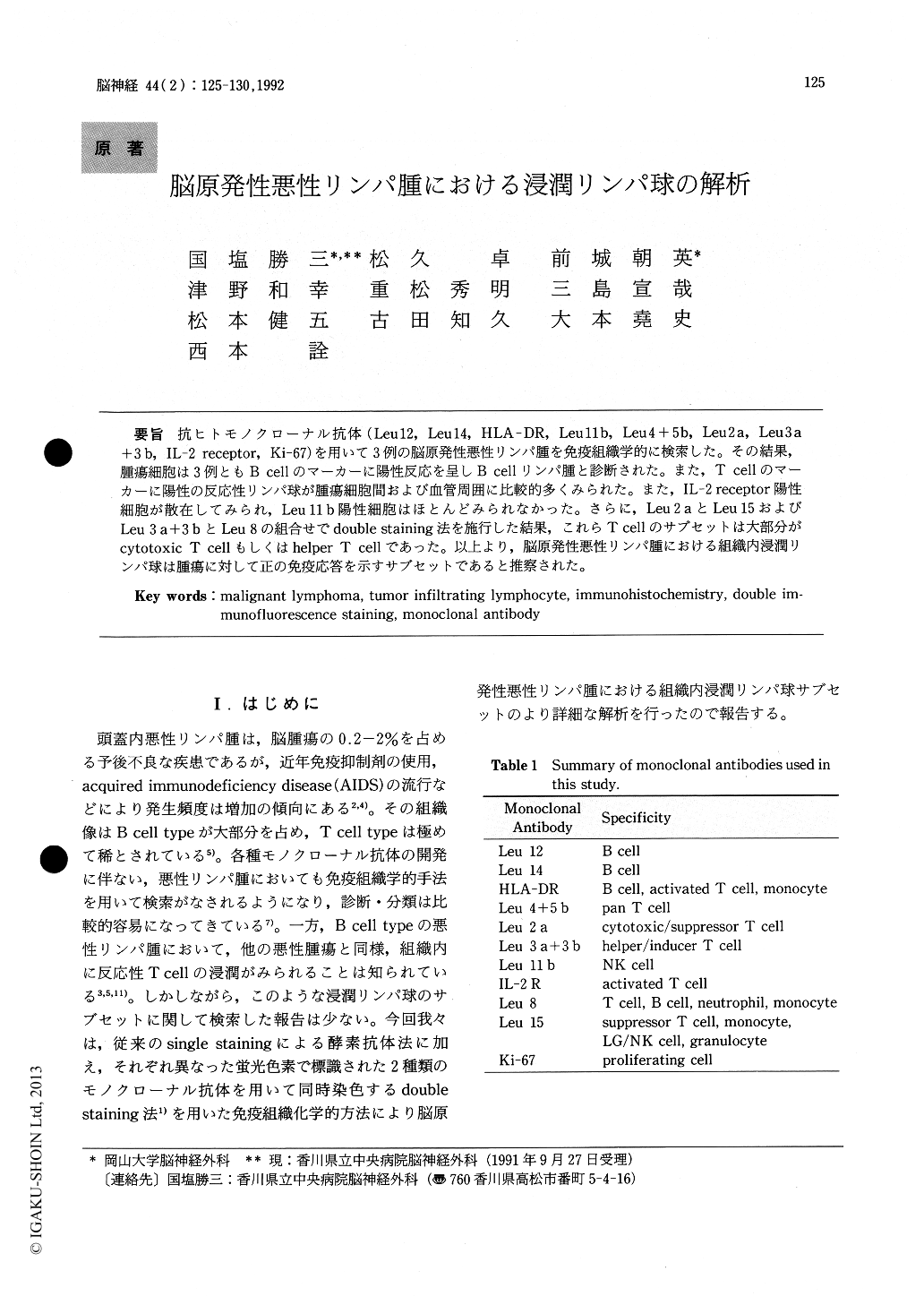
抗ヒトモノクローナル抗体(Leu 12, Leu 14, HLA-DR, Leu 11 b, Leu4+5b, Leu 2 a, Leu 3 a+3b, IL−2 receptor, Ki−67)を用いて3例の脳原発性悪性リンパ腫を免疫組織学的に検索した。その結果,腫瘍細胞は3例ともB cellのマーカーに陽性反応を呈しB cellリンパ腫と診断された。また,T cellのマーカーに陽性の反応性リンパ球が腫瘍細胞間および血管周囲に比較的多くみられた。また,IL−2 receptor陽性細胞が散在してみられ,Leu 11 b陽性細胞はほとんどみられなかった。さらに,Leu 2 aとLeu 15およびLeu 3 a+3bとLeu 8の組合せでdouble staining法を施行した結果,これらTcellのサブセットは大部分がcytotoxic T cellもしくはhelper T cellであった。以上より,脳原発性悪性リンパ腫における組織内浸潤リンパ球は腫瘍に対して正の免疫応答を示すサブセットであると推察された。
Three cases of malignant lymphoma were examined immunohistochemically using anti-Leu monoclonal antibodies (MAB) against B lympho-cytes, T lymphocytes subsets, NK cells, IL-2 recep-tor. In all three cases tumor cells were stained with anti-B cell's MABs, that is they were diagnosed as B cell type of malignant lymphoma. Moderate tumor-infiltrating lymphocytes in the parenchyma or aro-und blood vessels were observed, and majority ofThese results suggested that tumor-infiltrating lymphocytes in the malignant lymphoma weremainly activated cytotoxic or helper T cells.
these cells were T lymphocytes, demonstrating Leu 2a or Leu 3a + 3b positive phenotypes. IL-2 rece-ptor positive cells also found in the parenchyma of these tumors.
T lymphocyte subsets, such as Leu 2a positive cells and Leu 3a + 3b positive cells, were analyzed by means of double immunofluorescence staining method using the combination of Leu 2a and Leu15, or Leu 3a +3b and Leu 8 MABs. This method demonstrated that most of Leu 2a positive cells were negative for Leu 15 and that the majority of Leu 3a +3b positive cells were negative for Leu 8.These results suggested that tumor-infiltrating lymphocytes in the malignant lymphoma were mainly activated cytotoxic or helper T cells.

Copyright © 1992, Igaku-Shoin Ltd. All rights reserved.


