Japanese
English
- 有料閲覧
- Abstract 文献概要
- 1ページ目 Look Inside
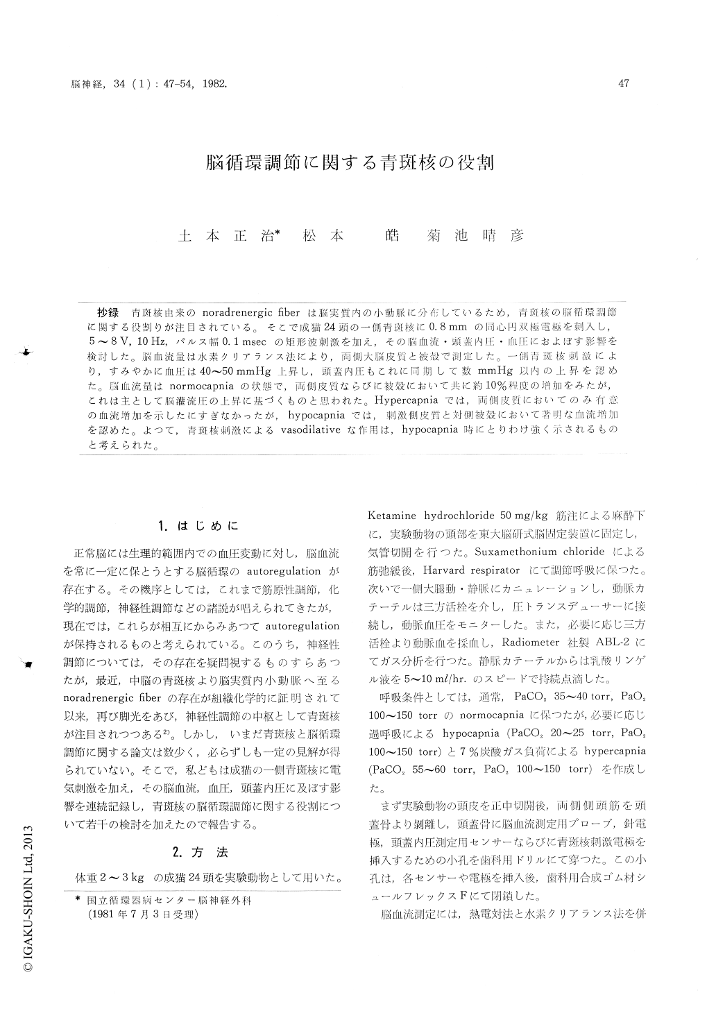
抄録 青斑核由来のnoradrenergic fiberは脳実質内の小動脈に分布しているため,青斑核の脳循環調節に関する役割りが注目されている。そこで成猫24頭の一側青斑核に0.8mmの同心円双極電極を刺入し,5〜8V,10Hz,パルス幅0.1msecの矩形波刺激を加え,その脳血流・頭蓋内圧・血圧におよぼす影響を検討した。脳血流量は水素クリアランス法により,両側大脳皮質と被殻で測定した。一側青斑核刺激により,すみやかに血圧は40〜50mmHg上昇し,頭蓋内圧もこれに同期して数mmHg以内の上昇を認めた。脳血流量はnormocapniaの状態で,両側皮質ならびに被殻において共に約10%程度の増加をみたが,これは主として脳灌流圧の上昇に基づくものと思われた。Hypercapniaでは,両側皮質においてのみ有意の血流増加を示したにすぎなかったが,hypocapniaでは,刺激側皮質と対側被殻において著明な血流増加を認めた。よつて,青斑核刺激によるvasodilativeな作用は,hypocapnia時にとりわけ強く示されるものと考えられた。
Recent histochemical studies suggested noradre-nergic fibers possibly originating from the locus coeruleus were distributed over the intracerebral arterioles. From this reason, locus coeruleus has been highlighted as a neurogenic control center of cerebral circulation. Several papers has been published concerning locus coeruleus and regulation of cerebral circulation, but still there is no agreed opinion for this problem. In the present study, we investigated the role of locus coeruleus on the regulation of cerebral circulation in cats, by record-ing the effects of electric stimulation of the uni-lateral locus coeruleus on cerebral blood flow, arterial blood pressure and intracranial pressure in normocapnic, hypocapnic and hypercapnic condi-tions.
Twenty four cats were anesthetized with keta-mine hydrochloride and placed in a stereotaxic instrument. These animals were also immobilized with suxamethonium chloride and placed under mechanical ventilation with room air. Catheterswere inserted into a femoral vein for infusion of lactated Ringer's solution, and a femoral artery for sampling of arterial blood and monitoring arterial blood pressure. Cerebral blood flow were measured by not only heat clearance method, but also hydro-gen clearance method. In hydrogen clearance method, four platinum electrodes were inserted into bilateral cortices of the middle ectosylvian gyrus and putamens. Intracranial pressure were measured by an epidural pressure sensor continu-ously. A concentric type bipolar electrode was inserted into unilateral locus coeruleus stereotaxi-cally and rectangular electric stimulation (5-8 volts, 10 Hz, pulse width O.1 msec.) was done at need.
Electric stimulation of unilateral locus coeruleus caused a rise of arterial blood pressure of 40-50 mmHg in all (normocapnic, hypocapnic and hyper-capnic) conditions. In normocapnia, bilateral cortical and putaminal blood flows increased up to 110% of control (non-stimulated) values during stimula-tion on locus coeruleus. In hypercapnic state, the changes of cerebral blood flow were not so much increased as in normocapnia. However in hypocapnia, ipsilateral cortical and contralateral putaminal blood flows extremly increased up to 152.8% and 138.4% of control values respectively following unilateral stimulation. During stimula-tion on locus coeruleus, intracranial pressure elevated within a few mmHg, synchronized with the elevation of blood pressure. When this stimu-lation stopped, the increased intracranial pressure returned to the prestimulation level immediately. But after the stimulation on locus coeruleus, in-tracranial pressure increased gradually and progres-sively with time, and finally reached up to about 30 mmHg in 11 out of 19 experimental animals.
From these results. it is concluded that electric stimulation of unilateral locus coeruleus caused vasodilatation on the cerebral arterioles, especially in hypocapnic condition.

Copyright © 1982, Igaku-Shoin Ltd. All rights reserved.


