Japanese
English
- 有料閲覧
- Abstract 文献概要
- 1ページ目 Look Inside
I.はじめに
環境衛生・予防衛生の改善と化学療法の進歩・普及に伴つて,脳結核腫は開発途上国を除いてはまれにしか見られなくなつた。わが国でも,ここ20〜30年来臨床医が脳結核腫に接することはきわめてまれであり,したがつて,器質性頭蓋内疾患の鑑別診断に際しても,ともすればはじめから結核腫の可能性は念頭にもおかないことが多い。しかし,脳結核腫が,今日もなお,わが国においても10),欧米諸国においても1),散発性にではあるが存在することも事実であり,この"potentially curable"な疾患の早期の診断と適切な治療は決してその意義を失つたわけではない。
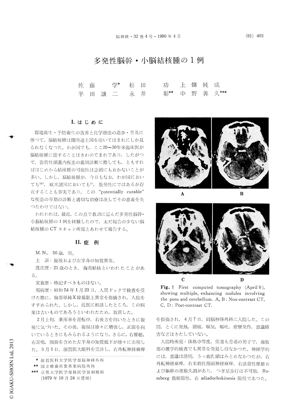
われわれは,最近,この点で教訓に富んだ多発性脳幹・小脳結核腫の1例を経験したので,未だ報告の少ない脳結核腫のCTスキャン所見とあわせて報告する。
A case of multiple cerebral tuberculomata in-volving the pons and cerebellum was presented. The lesions were demonstrated by CT as isodense to slightly dense foci. All four intra-axial lesions showed homogeneous enhancement following an intravenous injection of the contrast medium, and one of them was surrounded by a small area of low density, probably representing the perifocal edema.
The patient responded well to chemotherapy with streptomycin, hydrazid and rifampicin: cranial nerve signs and long tract signs clearing rapidly and the enhancing lesions and mass effect on CT disappearing concomitantly.
Although cerebral tuberculoma is nowadays very rare in Japan, still a high index of suspicion should always be entertained during the investigation of patients showing solitary or multiple enhancing lesions with no or slight degree of perifocal edema on CT, and a trial of antituberculous drugs should be given before the incurable malignancy is pre-sumed or the lesion is explored surgically.

Copyright © 1980, Igaku-Shoin Ltd. All rights reserved.


