Japanese
English
- 有料閲覧
- Abstract 文献概要
- 1ページ目 Look Inside
I.はじめに
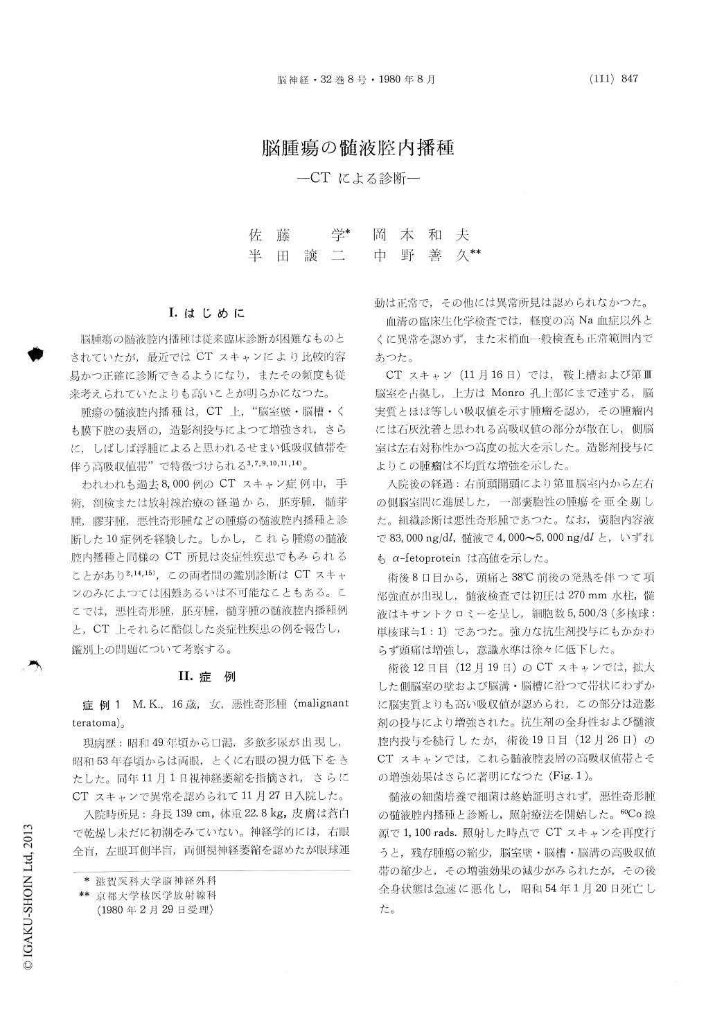
脳腫瘍の髄液腔内播種は従来臨床診断が困難なものとされていたが,最近ではCTスキャンにより比較的容易かつ正確に診断できるようになり,またその頻度も従来考えられていたよりも高いことが明らかになつた。
腫瘍の髄液腔内播種は,CT上,"脳室壁・脳槽・くも膜下腔の表層の,造影剤投与によつて増強され,さらに,しばしば浮腫によると思われるせまい低吸収値帯を伴う高吸収値帯"で特徴づけられる3,7,9,10,11,14)。
Of 8000 consecutive patients studied with com-puted tomography, 10 patients with primary intracranial tumors (germinoma, medulloblastoma, malignant teratoma and glioblastoma) showed ven-tricular or leptomeningeal spread of the tumor cells.
In patients with leptomeningeal spread, computed tomography showed obliteration of basal cisterns and sulci with isodense or slightly hyperdense mass, which was markedly enhanced following administ-ration of the contrast medium. In cases of ventri-cular spread, a narrow zone of high density was noted on the ependymal surface, and it was alsomarkedly enhanced with the contrast medium.
Similar CT scan appearance of contrast enhance-ment in the subarachnoid space or the ventricular surface was, however, noted also in the infectious processes such as basal arachnoiditis or ependymitis,and the differentiation of the neoplastic process from the infectious lesions seemed impossible based on the CT scan appearance alone.

Copyright © 1980, Igaku-Shoin Ltd. All rights reserved.


