Japanese
English
- 有料閲覧
- Abstract 文献概要
- 1ページ目 Look Inside
I.はじめに
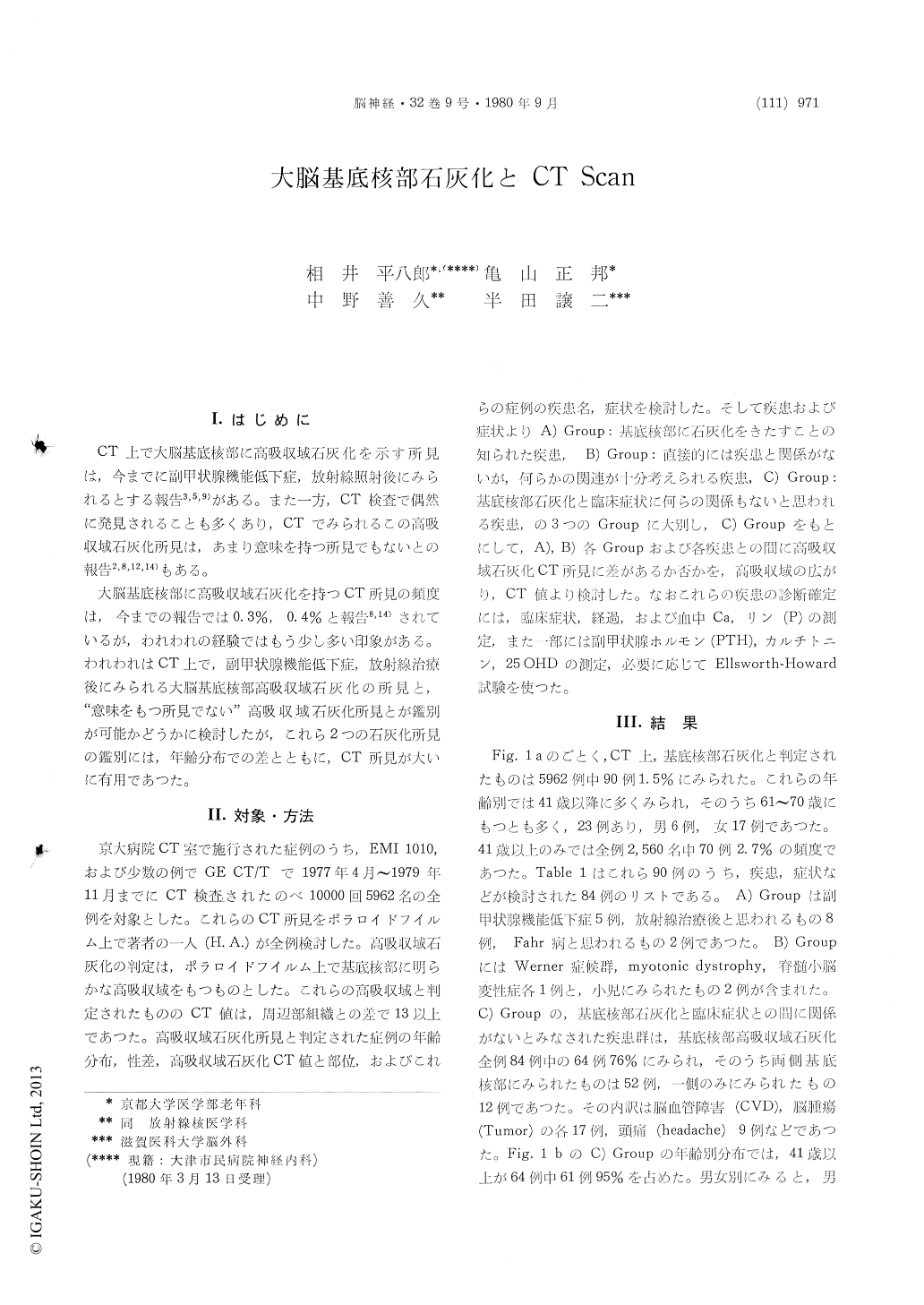
CT上で大脳基底核部に高吸収域石灰化を示す所見は,今までに副甲状腺機能低下症,放射線照射後にみられるとする報告3,5,9)がある。また一方,CT検査で偶然に発見されることも多くあり,CTでみられるこの高吸収域石灰化所見は,あまり意味を持つ所見でもないとの報告2,8,12,14)もある。
大脳基底核部に高吸収域石灰化を持つCT所見の頻度は,今までの報告では0.3%,0.4%と報告8,14)されているが,われわれの経験ではもう少し多い印象がある。われわれはCT上で,副甲状腺機能低下症,放射線治療後にみられる大脳基底核部高吸収域石灰化の所見と,"意味をもつ所見でない"高吸収域石灰化所見とが鑑別が可能かどうかに検討したが,これら2つの石灰化所見の鑑別には,年齢分布での差とともに,CT所見が人いに有用であつた。
A review of CT scans in consecutive 5962 patients revealed high density foci indicating calci-fication in the basal ganglia in 90 patients (1.6%). Six patients with incomplete clinical data were excluded, and the remaining 84 patients were divided into three groups.
Group A comprises 15 patients with conditions known to have calcification in the basal ganglia. Clinical diagnosis in this group was hypoparathyroid disease in 5, postirradiation in 8, and idiopathic brain calcification (Fahr's disease) in the remaining 2.
In group B, clinical condition in 5 patients were thought to have some relation, though not es-tablished yet, with calcification of the basal ganglia. One patient each with Werner's syndrome, myotonic dystrophy and spinocerebellar degeneration, and two children with mental retardation belonged to this group.
In 64 patients in group C, calcification in the basal ganglia was thought to be purely incidental. Clinical diagnosis in this group was cerebrovascular accident (CVA) in 17, brain tumor in 17, headache in 9, dementia of psychosis in 7, and miscellaneous in the remaining 14. Sixty-one out of 64 patients were 41 years of age or older. Two patients showed extrapyramidal symptoms and signs but the calcification in both patients seemed to be unrelated to their symptomatology.
Nonspecific, incidental calcification in this group C was characteristically confined to a focal area ofthe globus pallidus. This CT finding seems to conform well with the previous pathologic reports in which the oral part of the globus pallidus has been described as the site of predilection of non-specific microscopic calcification in the elderly. Attenuation of the dense focus in this group ex-ceeded that of the normal brain by 13 to 40 in CT number.
In comparison, computed tomographic calcification in patients with severe hypoparathyroid disease involved head of the caudate nucleus, putamen, dentate nucleus, cerebral cortex and cerebral white matter in addition to the globus pallidus, and the attenuation within the focus was much higher than that of group C. In patients with post-irradiation, location of the computed tomographic calcification was essentially similar to that in group C, but in some cases high density was noted diffusely in the basal ganglia.
In group B, patients presented with protean neurologic symptoms and signs, such as extra-pyramidal manifestations, convulsive seizures, various EEG abnormalities, mental retardation. In two children with mental retardation, calcification was noted also in the deep white matter. Cataracts are often seen not only in hypoparathyroid disease but also in Werner's syndrome and myotonic dys-trophy, and the patient with spinocerebellar de-generation in this series also showed cataracts. In this group, the presence of the some unidentified metabolic disorders is probable.
In conclusion, the possibility of specific disease showing calcification in the basal ganglia seems to be seriously considered, when:
(1) calcification in the basal ganglia is found in patients aged 40 or younger,
(2) attenuation of the dense focus exceeds that of the normal brain by more than 40 in CT number, or
(3) calcification involves caudate nucleus, putamen, dentate nucleus, cerebral cortex or cere-bral white matter.

Copyright © 1980, Igaku-Shoin Ltd. All rights reserved.


