Japanese
English
- 有料閲覧
- Abstract 文献概要
- 1ページ目 Look Inside
I.はじめに
内頸動脈閉塞症では,外頸動脈領域より眼動脈を介した側副血行にしばしば遭遇する。この場合,側副血行となつた眼動脈血流は"逆流"しており,この血流は方向指示型超音波血流計で,異常血流方向として検出できる。この原理を利用することにより,内頸動脈閉塞や高度狭窄を非観血的に診断可能とする報告は多い4,5,10〜12,17)。しかしながら,この側副血行の"有無"や"程度"に関する超音波血波検査所見の信頼性は充分検討されていない。
一方,内頸動脈閉塞症の臨床症状の多様性は,多く側副血行の有無や程度によるとされている2,3,21)ため,本症における側副血行の有無や程度の評価は臨床的にことに重要である。この側副血行の評価は,現在のところ脳血管撮影によらざるを得ず,本検査の副作用を考慮すると,頻回かつ経時的に施行することはちゅうちょされる。したがつて,非観血的検査法である超音波血流検査は注目されており,本法によるこの眼動脈側副血行の有無と程度の評価は,臨床的重要性を有する。
Ultrasonic Doppler technique as a diagnostic toolfor internal carotid arterial occlusions has beenintensively studied using the detection of ophthalmicblood flow directions. Nowadays, this technique isadmitted to have clinical usefulness. But the col-lateral flow via ophthalmic artery in internalcarotid arterial occlusions has not been quantit-atively evaluated by ultrasonic Doppler technique.
On 21 completely occluded internal carotid arteriesin 19 patients, the collateral flow via ophthalmicartery was evaluated both by ultrasonic Dopplertechnique on ophthalmic artery (ophthalmic arteryDoppler) and by carotid angiography.
Angiographical findings of the collateral flowthrough ophthalmic artery were graded in threedegrees according to the extent of this collateralflow to middle cerebral artery. It was expressedas "good collateralal" when the branches andstem of middle cerebral artery were filled throughthe ophthalmic collateral flow. "Poor collateral"was expressed in the cases in which only stem ofmiddle cerebral artery was showed. "No collateral"indicated no collateral flow via ophthalmic arteryon angiograms. There were good collateral in 6vessels, poor collateral in 2 vessels and no collateralin 13 vessels.
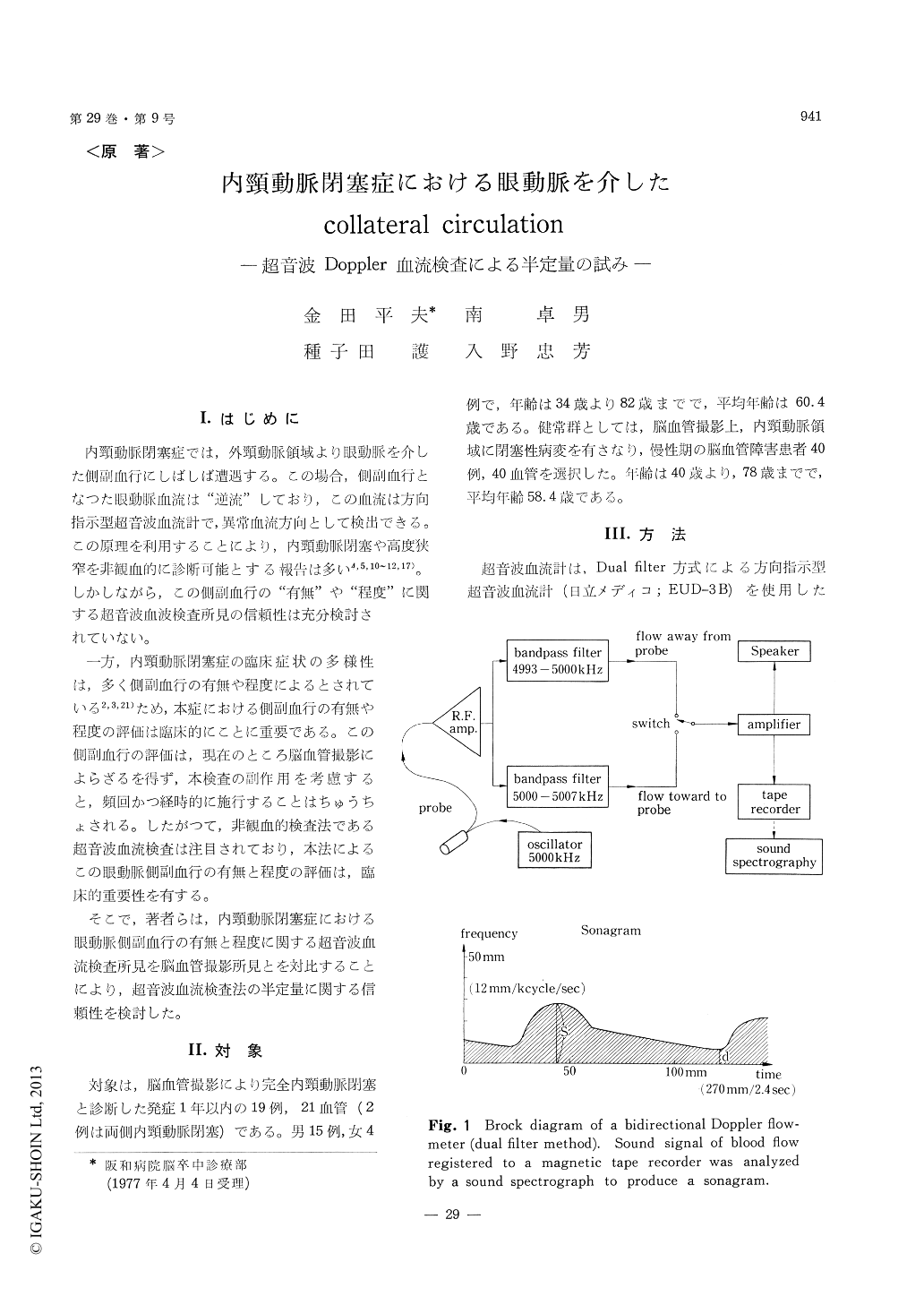
Using a directional ultrasonic Doppler flowmeter(dual filter method) and a sound spectrograph, theophthalmic blood flow signal was obtained as asound spectrogram (sonagram). In sonagrams, thedistance to the point of maximum blood flowvelocity during the systolic phase from the baseline was expressed as "S" (mm; 12 mm=1 kcycle/sec). The findings of the ophthalmic artery Dop-pler were classified in three types; physiologicalflow type, no flow signal type and reversed flowtype. Moreover, the reversed flow type was gradedin three patterns according to the degree of "S".We defined that a sonagram of S>40 mm was"severe reversed flow pattern", that of 40 mm≧S≧20 mm was "moderate reversed flow pattern"and that of S<20 mm was "poor reversed flowpattern". There were five vessels of severe reversedflow pattern, three vessels of moderate reversedflow pattern and five vessels of poor reversed flowpattern. No flow signal type in four vessels andphysiological flow type in four vessels were alsoexisted.
In five vessels of severe reversed flow pattern,all revealed good collaterals on angiograms. Inthree vessels of moderate reversed flow pattern,two of them were poor collaterals. And in theother thirteen vessels of poor reversed flow pattern,no flow signal type and physiological flow type,all the vessels except for one had no collateralsangiographically.
Therefore, in 21 occluded internal carotidarteries except for two vessels, the findings of theophthalmic artery Doppler well correlated to theangiographical findings. In two non-correlatedvessels, there existed definite causes i. e. the angio-graphical technical factor and the time lag betweenangiography and ultrasonic Doppler examination.
Conclusively, the ophthalmic artery Doppler hasclinical usefulness not only in diagnosis but alsoin semi-quantitative evaluation of the ophthalmiccollateral flow in internal carotid arterial occlusions.

Copyright © 1977, Igaku-Shoin Ltd. All rights reserved.


