Japanese
English
- 有料閲覧
- Abstract 文献概要
- 1ページ目 Look Inside
I.はじめに
視路は網膜にはじまり視神経,視交叉,外側膝状体,視放線を経て後頭葉視覚領に至る長い経路を有し,あたかも頭蓋内に広くアンテナを張りめぐらしているようなもので,もし頭蓋内のどこかに病変が生じると,このアンテナに大なり小なり把えられ,その視路障害のpatternより頭蓋内病変の局在を知ることができる。従つて頭蓋内病変の診断にあたつて視路障害の有無を検討することは極めて重要である。
一般に視路障害の検索には,視力・視野検査など患者の供述に頼る検査が行なわれているが,実際脳神経外科で扱う患者のなかには,意識障害,失語症などのため意志疎通が充分期待できない場合も少なくない。
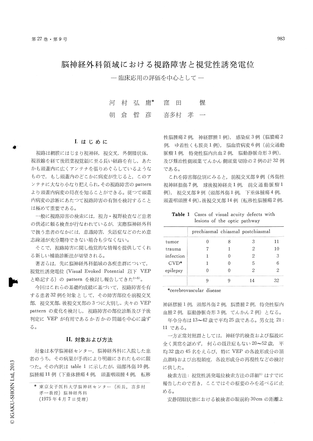
著者らは,先に脳神経外科領域の各疾患群について,視覚性誘発電位(Visual Evoked Potential以下VEPと略記する)のpatternを検討し報告してきた1〜5)。 今回はこれらの基礎的成績に基づいて,視路障害を有する患者32例を対象として,その障害部位を前視交叉部,視交叉部,後視交叉部の3つに大別し,夫々のVEPpatternの変化を検討し,視路障害の部位診断及び予後判定にVEPが有用であるか否かの問題を中心に論ずる。
The present study attempted to evaluate theclinical usefulness of the VEP technique for assesingvisual field defects and its prognosis in the field ofneurological surgery.
Visual evoked potential (VEP) was studied in 45normal subjects and 32 patients with visual defectdue to lesion of the optic pathways, i. e. lesion onprechiasmal, chiasmal and potchiasmal level re-spectively.
Patient lying with closed eyes on a bed in thedark room was stimulated with a sequence of flashesfrom a stroboscope placed 30-40 cm in front of thesubject's and flickered by a photic stimulator.Stoboscope was flashed on both eyes simultaneously,and on right and left eye separately. Fifty repeatedevoked responses were avaraged by means of ATAC201 type computer with binocuar and monocularstimulation.
Effects of visual pathway lesion upon VEPs wereas follows;
1) VEP in cases with prechiasmal lesion (9 cases):In all of 9 cases (100%), VEPs obtained by stimu-lating the affected eye were characterized by thepresence of marked suppression or larking of eachcomponent bilaterally. On the contrary, VEPsappeared bilateral symmetrical pattern, when botheyes and normal eye were stimulated respectively(fig. 4 or upper in fig. 9).
2) VEP in cases with chiasmal lesion (9 cases):In only 3 out of 9 cases (33%) with typical bi-temporal hemianopia, VEPs revealed symmetricalpattern in response to binocualr stimulation. VEPsobtained by monocualr stimulation appeared muchlarger response in the ipsilateral side to the stimu-lated eye than that in the contralateral side (fig. 5).
In 6 out of 9 cases (67%), VEPs showed variouspattern without above mentioned findings as fig. 5,because chiasmal lesions caused by traumal, vasculardisorders, brain tumors, etc. sometimes extendednot only to chiasmal but also prechiasmal or post-chiasmal region (fig. 6).
3) VEP in cases with postchiasmal lesion (14 cases): 8 out of 14 (57%) with homonymous hem-ianopia had a symmetrical pattern of VEPs obtainedon binocular stimulation. On the other hand, VEPswith monocular stimultiaon showed marked asym-metry of suppression or lacking of VEP on theaffected hemisphere in 12 out of 14 cases (85.7%)(fig. 8).
4) There was the intimate relation between im-provement of visual symptoms and alteration ofVEPs. On conclusion, so far as the locarizationand prognosis of visual disorder are concerned, theVEP may be useful as a diagnostic aid in thefield of neurological surgery.

Copyright © 1975, Igaku-Shoin Ltd. All rights reserved.


