Japanese
English
- 有料閲覧
- Abstract 文献概要
- 1ページ目 Look Inside
はじめに
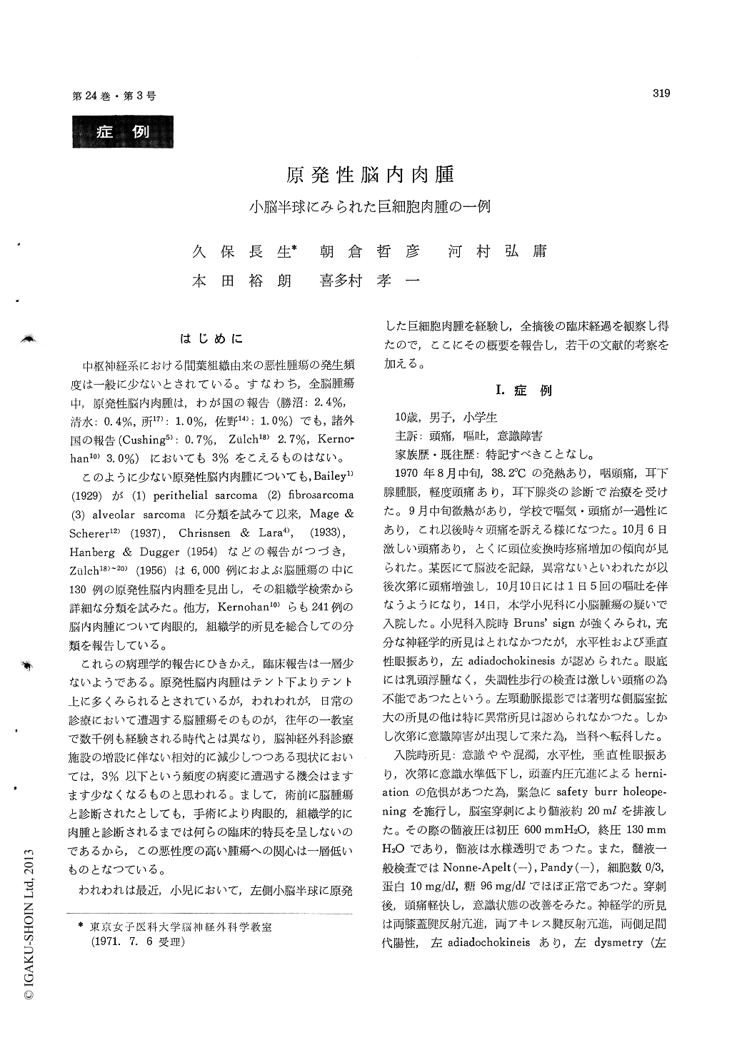
中枢神経系における間葉組織由来の悪性腫瘍の発生頻度は一般に少ないとされている。すなわち,全脳腫瘍中,原発性脳内肉腫は,わが国の報告(勝沼:2.4%,清水:0.4%,所17):1.0%,佐野14):1.0%)でも,諸外国の報告(Cushing5):0.7%,Zulch18)2.7%,Kerno—hanio)3.0%)においても3%をこえるものはない。
このように少ない原発性脳内肉腫についても,Bailey1)(1929)が(1) perithelial sarcoma (2) fibrosarcoma(3) aiveolar sarcomaに分類を試みて以来,Mage &Scherer12)(1937),Chrisnsen & Lara4),(1933),Hanberg & Dugger (1954)などの報告がつづき,Zülch18)〜20)(1956)は6,000例におよぶ脳腫瘍の中に130例の原発性脳内肉腫を見出し,その組織学検索から詳細な分類を試みた。他方,Kernohan10)らも241例の脳内肉腫について肉眼的,組織学的所見を総合しての分類を報告している。
From a pathogenetic point of view, the existence of intracranial giant cell sarcoma is still contro-versial and there is disagreement of several opin-ions. However, it is a fact that many investigators have attempted to confirm the presence of intra-cranial giant cell sarcoma and to classify the signi-ficance of the tumor. In our clinic, recently a case of suspicious giant cell sarcoma of the cere-bellar hemisphere was experienced. The patient, 10 year old boy had revealed only signs of intra-cranial hypertension and the left cerebellar hemi-sphere, but no significant different signs from those of other cerebellar neoplasms such as a astrocytoma or medulloblastoma, except for a rush of clinical course. A tumor of the left cerebellar hemisphere identified by vertebral angiography and iodoventriculography had been surgically ex-tirpated without any events. Microscopic findings of the tumor were as follow in H-E and silver stains : The cell pattern was mainly composed of spindle shaped and lymphoid ones. Between these were frequent appearance of bizzarre giant cells containing polymorphic nuclei. The reticulum was not only limited within perivascular areas, but also scattered widely spreading throughout the neoplasmic tissue. A histological diagnosis of giant cell sarcoma was proposed, however, the confirm-ative evidences seemed to be unsatisfactory for a final decision. Five months after the first surgery, the patient complained of sudden occurrence of dyspnea and tetraplegia. Emergency laminectomy between C 2-C 7 revealed extensive metastasis of the tumor in the extra- and intramedullary spaces of the cervical cord. Histological findings of the cervical cord tumor was almost similar to those of the cerebellar tumor. Electron microscopic study revealed the presence of numerous filaments (100 Å-150 Å in width) within the cytoplasm of giant cells. The nature of the filaments in still obscure, and it is unable to differentiate them from collagen fibers. It might be considered that giant cell sar-coma could be exist intracranially, however, further investigation should be necessary so as to clarify the problem concerning the pathology of this tumor.

Copyright © 1972, Igaku-Shoin Ltd. All rights reserved.


