Japanese
English
- 有料閲覧
- Abstract 文献概要
- 1ページ目 Look Inside
I.はじめに
幼児において,それまで全く健康であつたものが,突然痙攣発作と意識障害を起こし,その後に片麻痺を残す,いわゆるAcute infantile hemiplegiaの内,近年の血管撮影の発達により血管異常の証明されるものが増加してきている。しかし,いまだに原因不明と考えられるものが多いのが現状であり13),かつ,急性期で死亡することが少なく,急性期の神経病理学的検討はほとんどみられていない3)。最近,私達は不幸にして急性期に死亡したAcute infantile hemiplegiaの1症例を経験したので,その病理所見を報告するとともに,発生原因に関する考察を試みた。
The recent applications of cerebral angiographyfor the investigation of acute infantile hemiplegiadisclosed various kinds of vascular occlusive diseasesas the causes of this condition. However, still manycases fall into the category of acute infantile he-miplegia of obscure origin. In addition, very fewcareful post-mortem examinations in acute stagehave been documented in the literature.
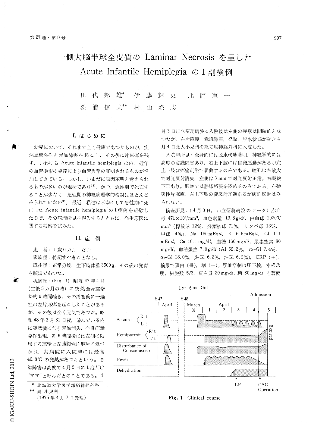
A 1 year 6 months old girl with a past historyof transient left hemiplegia at 5 months of agesuddenly developed stupor and generalized convul-sion, followed 6 hours later by left-sided seizuresand left flaccid hemiplegia. The patient was ad-mitted to Hokkaido University Hospital 4 days laterin poor general conditions.
Neurological examinations revealed stuporousgirl with dilated right pupil, and left hemiplegia.Right carotid arteriogram showed a shift of anteriorcerebral artery to the left and questionable avas-cular area in the right occipital region (Fig. 2).
The patient went into a shock during emergencyoperation under the presumptive diagnosis of acutesubdural hematoma, subsequently developed respira-tory distress, and expired on the 5th day from theonset.
Autopsy was limited to the brain. The cerebralhemispheres were generally swollen, with cerebel-lar herniation. Coronal sections of the cerebralhemispheres disclosed a shift of the mid-line struc-tures to the left, and prominent laminar necrosisat the deeper layers of right entire cortex (Fig. 3,& 4).
Histologically, the laminar necrosis mainly in-volved the IIIrd, IVth and Vth layers, and showedslight sponginess, loss of neurons and oligodendro-glia, and eosinophilic neuronal changes, withoutastroglial or microglial proliferations (Fig. 5).However, both neuronal loss and gliosis were foundin the end plate and Sommer's sector of the righthippocampus, suggesting that these changes weredue to both present and previous episodes of he-miplegia (Fig. 6). The left hemisphere, bain stemand cerebellum were free from anoxic changes(Fig. 7), and bilateral carotid arteries at the circleof Willis were not remarkable.
The distribution of the cortical laminar necrosisclearly indicates the lesions were located in thedistribution of right anterior, middle and posteriorcerebral arterial territories without generalizedanoxic changes. Although no carotid lesions werevisualized by anteriogram or post-mortem examina-tion, the anatomical evidence in this case pointsto vascular process as the cause of acute infantilehemiplegia, the exact mechanism of which is notclear at present.
Copyright © 1975, Igaku-Shoin Ltd. All rights reserved.