Japanese
English
- 有料閲覧
- Abstract 文献概要
- 1ページ目 Look Inside
I.緒言
頭蓋内において脳を被包する脳膜三葉のうち,最も外側の硬脳膜がきわめて強靱であるのに反し,その内側のくも膜および軟脳膜はきわめて非薄であり,容易に離断されやすい。このため硬脳膜の最大の役割は,頭蓋発育期における内骨膜としての作用を除けば,脳を頭蓋骨内面から分離し,頭蓋腔内における脳移動時に脳表面を庇護するとともに,脳が頭蓋内面へうちつけられることによつて生ずる運動量およびこれによつてひき起こされる脳全体としての振動振幅に緩衝作用をなしている。このため開頭手術にあたつて,硬脳膜が切除されたり,退縮した場合にそのまま放置し硬脳膜が部分的欠損のままとされる場合には,脳表面が硬い頭蓋骨内面に直接に接触し脳表面が損傷されやすい状況におかれることになる。また脳表面のくも膜下腔は,開頭手術特に脳組織に直接侵襲を加えるような操作の場合には開放され,このためくも膜下腔を循環する髄液はこの部を経てくも膜下腔の外に流出する。他方,このような髄液漏出があれば,開頭手術部の整復骨片縁の骨間隙を経て髄液が頭皮下へも流出し,これが頭皮手術創縫合部の治癒をいちじるしく阻害する。またこの部を経て髄液が頭皮外へも流出する可能性が大となり,逆行性頭蓋内感染が行なわれることになる。このため開頭手術における髄液の頭蓋腔外漏出を防がねばならず,この目的には硬脳膜の完全閉鎖以外に方法はない。他方,硬脳膜欠損とともにその直下のくも膜,軟脳膜の損傷が同時に存在すれば,遊離硬脳膜縁と脳組織との間に結合織が産生されて,両者は瘢痕形成によつて接合され,これがてんかん発生の原因となりうる。
以上のような理由から,頭部外傷や開頭手術操作によつて硬脳膜に欠損部が生じた場合には,なんらかの方法によつてこれを補墳しておかねばならない。このような理由から硬脳膜欠損部に対する補垣材や補垣法については早くから研究が行なわれ,すでに1890年にはBeach1)によつて金属箔による硬脳膜欠損部補墳法が行なわれ,その後1895年Abee2)によるゴム片,1909年Kirschner3)による筋膜補墳法が行なわれた。その後現在にいたるまで,諸家により硬脳膜補墳材として各種の物質が実験的および臨床的に研究がなされ,臨床応用がなされてきている。しかし,いずれも硬脳膜補墳材としてもつべき諸条件を満足していない。このため,近年においてもなお,理想的硬脳膜補旗材の探索が行なわれている現状である。
The dural defect have to repaire by some kinds of material in craniotomy to protect the cortical surface from the skull while in moving of head, production of meningocerebral adhesion and leakage of cerebrospinal fluid from subarachnoid space.
Many research had done for the dural substitute by many neurosurgeons and no ideal one was ob-tained.
The authors had made the fundamental studies on the absorbable gelatin film by dog. The absorb-able gelatin film was transplanted in the area of artificial dural defect of dog. The gelatin film placed on the normal and mechanical damaged cortical surface and its edge is inserted between the dural edge and cortical surface.
The authors had emphasized that the transplanted absorbable gelatin film is replaced with new fibrous membrane after its absorption without production of meningocerebral adhesion.
The course of new membrane formation were divided into three stages as follows.
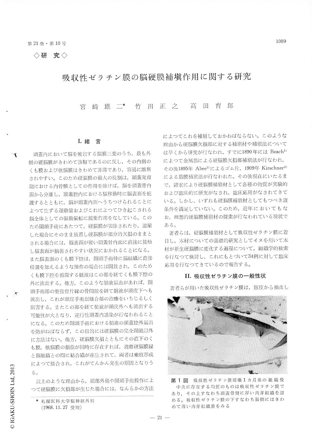
The first stage is that of reaction for the gelatin film and continue in initial one month. The outer surface of gelatin film is covered by layer of gra-nulation tissue at first and the inner surface of gelatin film also is covered by thin layer of gra-nulation tissue layer in latter,
The second stage is that of absorption of gelatin film for three months. The transplanted gelatin film shows fragmentation by foreign body giant cell and then absorpt gradually.
The third stage is that of production of fibrous tissue in granulation tissue after fifth month. The tranplanted gelatin film disappeared completely and the granulation tissue is replaced with fibrous tissue and new membrane is established.
The authors had also done the clinical research on the absorbable gelatin film. The gelatin film had transplanted in 34 cases and used in dural defect by external decompression in 17 cases, in dural lacera-tion by depressed fracture in 4 cases and dural defect by reced of dural edge in 6 cases of subtentorial operation etc.
The authors' detail technique for clinical use of absorbable gelatin film was described and the advan-tages and disadvantages was disussed also.

Copyright © 1969, Igaku-Shoin Ltd. All rights reserved.


