Japanese
English
- 有料閲覧
- Abstract 文献概要
- 1ページ目 Look Inside
I.緒言
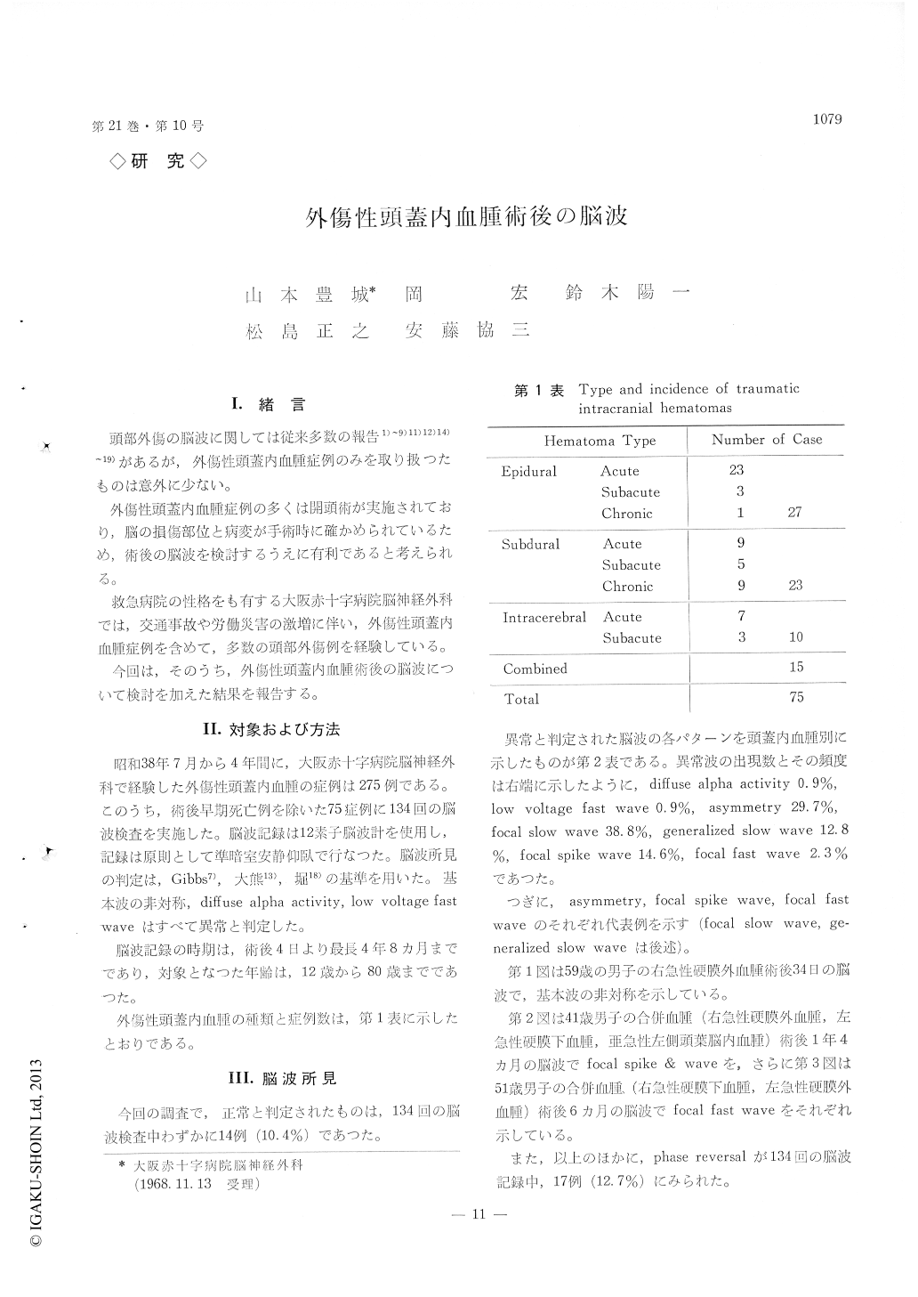
頭部外傷の脳波に関しては従来多数の報告1)〜9)11)12)14)〜19)があるが,外傷性頭蓋内血腫症例のみを取り扱つたものは意外に少ない。
外傷性頭蓋内血腫例の多くは開頭術が実施されており,脳の損傷部位と病変が手術時に確かめられているため,術後の脳波を検討するうえに有利であると考えられる。
For recent 4 years, 275 cases of traumatic intra-cranial hematomas were operated on at the Depart-ment of Neurosurgery, Osaka Red Cross Hospital.
Seventy-five cases of those hematomas, in which early death cases were excluded, were examined with successive 131 EEG records.
1) The period of EEG recording extended from 4 days to 4 years and 8 months after the operation. The range of age was 12 to 80 years. Through the whole records, the normality was found only 10. 4%.
2) The classification of abnormal EEG patterns and their incidences were as follows. Diffuse alpha 0.9%, low voltage fast 0.9%, asymmetry 29. 7%, focal slow 38. 8%, generalized slow 12. 8%, focal spike 14.6%, and focal fast 2.3%. Phase reversal phenomenon was seen in 12.7%.
3) Posttraumatic epilepsy occurred in 9 cases of this series (12%), and 8 cases of those developed epilepsies from 4 months to 2 years after head in-juries. All cases were severe in head injuries, showing early epilepsy, skull fracture and lobe syndrome. Cerebral contusion was found mainly in the frontal and temporal region and EEG revealed focalized abnormality. There was no remarkable relation between the forms of clinical epileptic sei-zure and EEG patterns.
4) Cerebral contusion may lead to focal lesion of meningo-cerebral cicatrix and/or cerebral cicatrix in convalescent period.
It is conceivable that a cause for posttraumatic epilepsy is not intracranial hematoma itself, but brain scar, which may form epileptogenic focus.
5) Since such severe head injury cases mentioned above are expected to have a higher possibility to develop posttraumatic epilepsy in future, follow-up study with EEG recording should be necessary.

Copyright © 1969, Igaku-Shoin Ltd. All rights reserved.


