Japanese
English
- 有料閲覧
- Abstract 文献概要
- 1ページ目 Look Inside
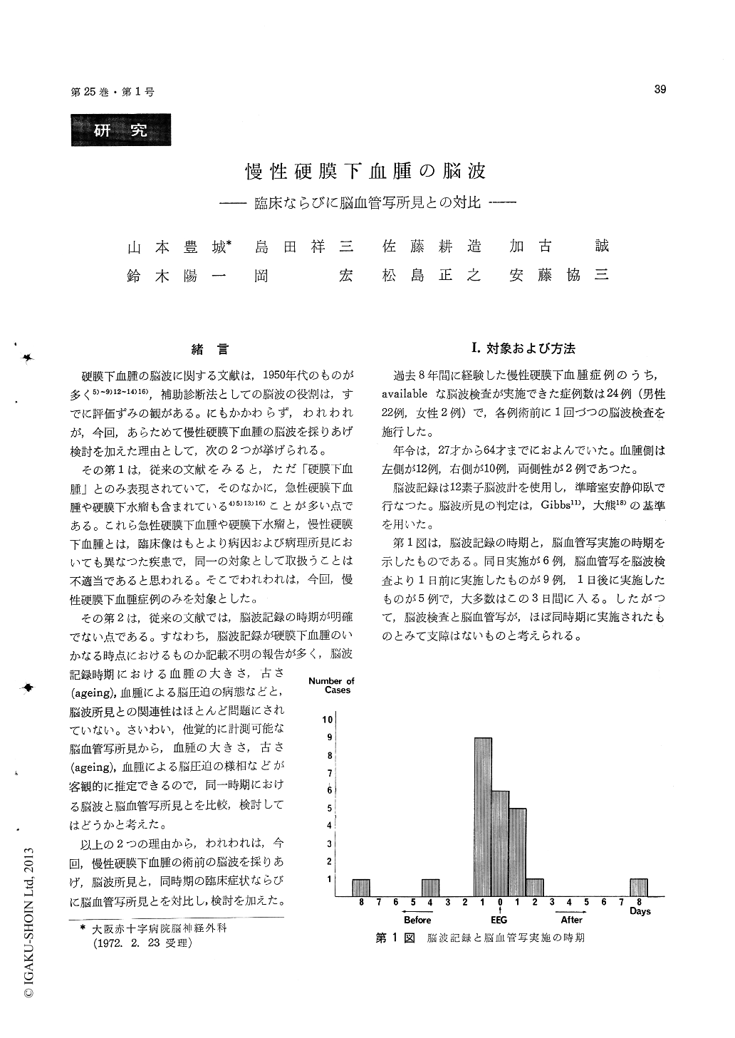
緒言
硬膜下血腫の脳波に関する文献は,1950年代のものが多く5)〜9)12〜14)16),補助診断法としての脳波の役割は,すでに評価ずみの観がある。にもかかわらず,われわれが,今回,あらためて慢性硬膜下血腫の脳波を採りあげ検討を加えた理由として,次の2つが挙げられる。
その第1は,従来の文献をみると,ただ「硬膜下血腫」とのみ表現されていて,そのなかに,急性硬膜下血腫や硬膜下水瘤も含まれている4)5)13)16)ことが多い点である。これら急性硬膜下血腫や硬膜下水瘤と,慢性硬膜下血腫とは,臨床像はもとより病因および病理所見においても異なつた疾患で,同一の対象として取扱うことは不適当であると思われる。そこでわれわれは,今回,慢性硬膜下血腫症例のみを対象とした。
Previous literatures on the electroencephalogram in the cases of subdural hematoma have, in the main, dealt with the type of electrical activity and with availability of EEG in localizing the lesion. However, few workers have taken up a problem between EEG findings and the size of the hema-toma, and between EEG findings and pathological brain changes caused by the hematoma.
In the present study, an attempt was made to correlate EEG findings in the cases of chronic subdural hematoma with the symptomatology and simultaneous cerebral angiographic findings.
Preoperative electroencephalographic studies were made on 24 patients (22 male and 2 female) with surgically proved chronic subdural hematoma. The range of age was 27 to 64 years. Left-sided hema-tomas were seen in 12 cases, right-sided in 10 cases and bilateral in 2 cases.
Through the whole records, normal EEG was found only in 3 cases (12.5%), borderline EEG was found in 1 case (4.2%) and abnormal EEG wasfound in 20 cases (83.3%).
Preoperative abnormal EEG was characterized by a marked reduction in amplitude of background activity in bipolar recording and a slow wave focus on the side of chronic subdural hematoma. Thus, abnormal EEG patterns could be classified as follows.
1) decreased amplitude of background activity over the site of the hematoma.
2) focal slow wave over the site of the hema-toma.
3) focal slow wave with decreased amplitude of background activity coincided with the site of the hematoma.
It can be pointed out that for the localization of the lesion, focal slow wave is more helpful than asymmetry of alpha or background activity, while the decreased amplitude of background activitymay sometimes lead to wrong lateralization.
EEG abnormality may correlate with neurologi-cal symptoms and signs such as headache, vomiting, disturbance of consciousness, pyramidal sign, papilledema and anisocoria.
EEG abnormality may also correlate with simul-taneous cerebral angiographic findings such as the type (size) of avascular area, shift of anterior cere-bral artery and inward displacement of angiographic Sylvian point in anteroposterior view.
Cerebral angiography is a more reliable and accurate test for demonstration of chronic subdural hematoma than other diagnostic studies. However, EEG is harmless and useful in screening test for the lesion.
Copyright © 1973, Igaku-Shoin Ltd. All rights reserved.