Japanese
English
- 有料閲覧
- Abstract 文献概要
- 1ページ目 Look Inside
I.はじめに
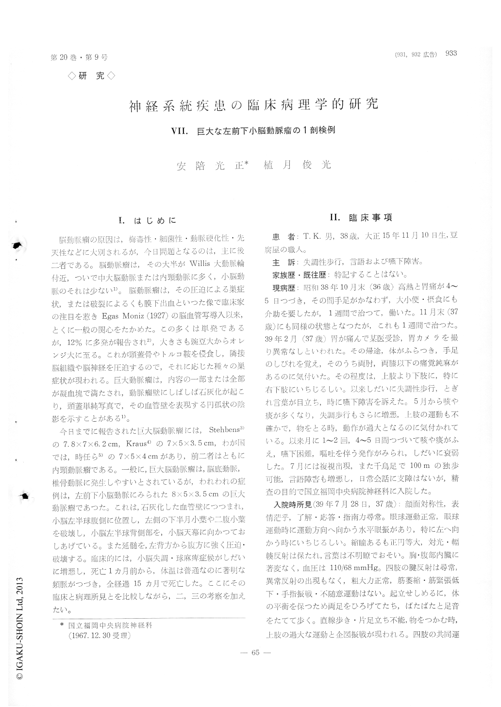
脳動脈瘤の原因は,梅毒性・細菌性・動脈硬化性・先天性などに大別されるが,今日問題となるのは,主に後二者である。脳動脈瘤は,その大半がWillis大動脈輪付近,ついで中大脳動脈または内頸動脈に多く,小脳動脈のそれは少くない1)。脳動脈瘤は、その圧迫による巣症状,または破裂によるくも膜下出血といつた像で臨床家の注目を惹きEgas Moniz (1927)の脳血管写導入以来,とくに一般の関心をたかめた。この多くは単発であるが,12%に多発が報告され2),大きさも豌豆大からオレンジ大に至る。これが頭蓋骨やトルコ鞍侵食し,隣接脳組織や脳神経を圧迫するので,それに応じた種々の巣症状が現われる。巨大動脈瘤は,内容の一部または全部が凝血塊で満たされ、動脈瘤壁にしばしば石灰化が起こり,頭蓋単純写真で,その血管壁を表現する円孤状の陰影を示すことがある1)。
今日までに報告された巨大脳動脈瘤には,Stehbens3)の7.8×7×6.2cm,Kraus4)の7×5×3.5cm,わが国では,時任ら5)の7×5×4cmがあり,前二者はともに内頸動脈瘤である。一般に,巨大脳動脈瘤は,脳底動脈,椎骨動脈に発生しやすいとされているが,われわれの症例は,左前下小脳動脈にみられた8×5×3.5cmの巨大動脈瘤であつた。これは,石灰化した血管壁につつまれ,小脳左半球腹側に位置し、左側の下半月小葉や二腹小葉を破壊し,小脳左半球背側部を,小悩天幕に向かつておしあげている。また延髄を,左背方から腹方に強く圧迫・破壊する。臨床的には,小脳失調・球麻痺症候がしだいに増悪し,死亡1カ月前から,体温は普通なのに著明な頻脈がつづき,全経過15ヵ月で死亡した。ここにその臨床と病理所見とを比較しながら,二,三の考察を加えたい。
A man, aged thirty-eight, developed the cerebellar ataxia and the bulbar palsy 15 months prior to his death which have gradually become worse, and he had a persistent tachycardia of about 120 per minute in spite of normal body temperature a month priorto his death. Finally he died of pneumonia.
The postmortem examination revealed the exis-tence of a large, children's fist-sized, tumor with a appearance similar to the teratoma at first glance, measuring 8×5×3.5 cm in size, in the ventral por-tion of the left cerebellar hemisphere, which was composed of a mass of small, round, conglomerate ones and which was covered by the calcified capsule. At close examination it was confirmed that the tumor is a congenital aneurysm derived from the left an-terior artery, the content of which consists of blood coagula ranging from fresh to old in estimated time. The aneurysm was situated at the ventral portion of the left cerebellar hemisphere, so that the left in-ferior semilunar and biventral lobules suffered severe damage. At the medulla the aneurysm produced the intense compression to the portions directing from dorsal toward ventral, resulting in the severe involvement of the nucleus dorsalis of nervus vague on both sides, the nucleus hypoglossus and the nu-cleus ambiguus especially on the left side.
In the present paper the author made some con-siderations on the relation between the pathological and clinical findings as described above.

Copyright © 1968, Igaku-Shoin Ltd. All rights reserved.


