Japanese
English
- 有料閲覧
- Abstract 文献概要
- 1ページ目 Look Inside
I.はじめに
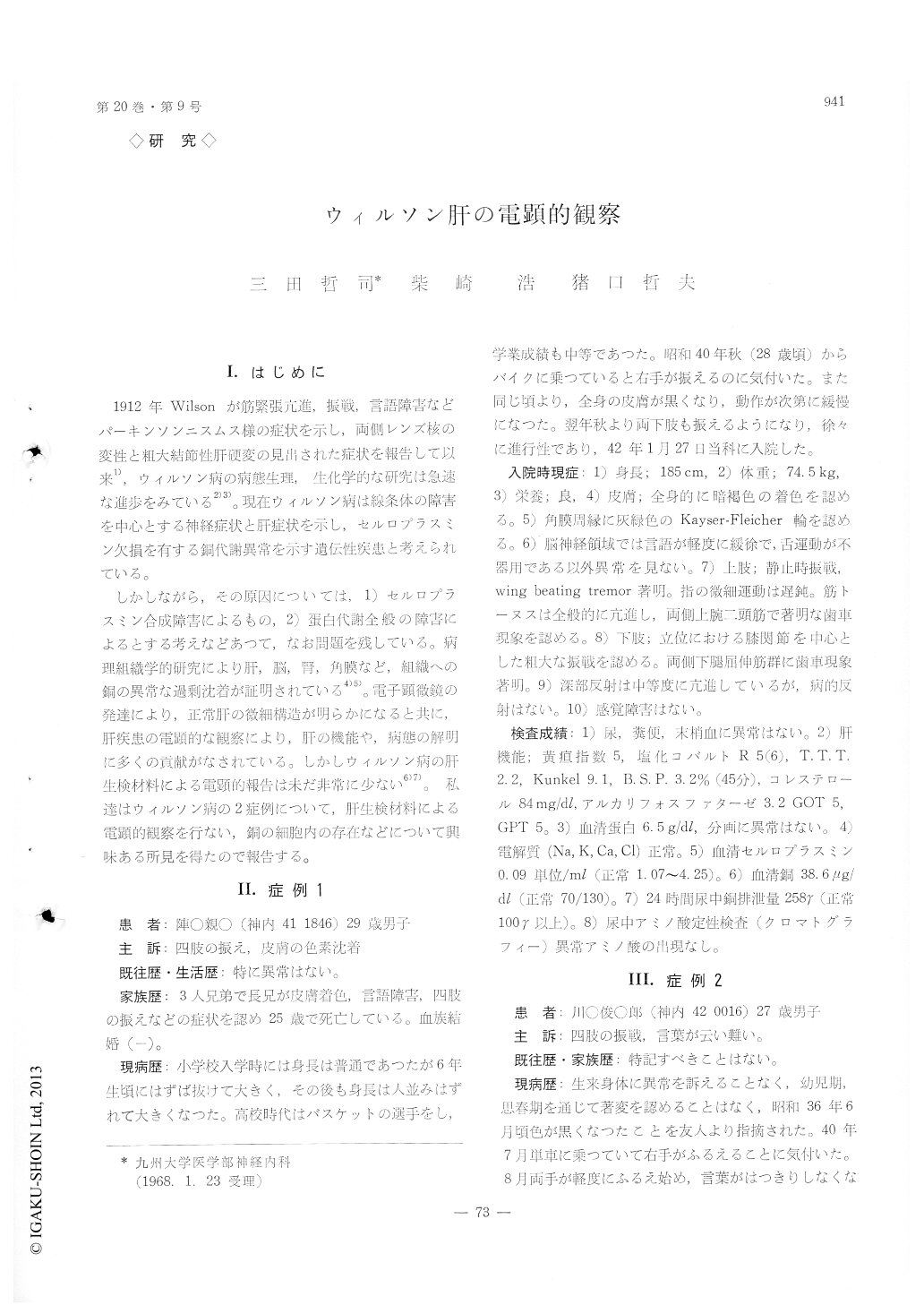
1912年Wilsonが筋緊張亢進,振戦,言語障害などパーキンソンニスムス様の症状を示し,両側レンズ核の変性と粗大結節性肝硬変の見出された症状を報告して以来1),ウィルソン病の病態生理,正化学的な研究は急速な進歩をみている2)3)。現在ウィルソン病は線条体の障害害を中心とする神経症状と肝症状を示し,セルロフラスミン久損を有す銅代謝異常を示す遺伝性疾患と考えられている。
しかしながら,その原因にっいては,1)セルロプラスミン合成障害によるもの,2)蛋白代謝全般の障害によるとする考えなどあつて,なお同題を残している。病理組織学的研究により肝,脳,腎,角膜など,組織への銅の異常な過剰沈着が証明されている4)5)。電子顕微鏡の発達により、正常肝の微細構造が明らかになると共に、肝疾患の電顕的な硬察により,肝の機能や,病態の解明に多くの貢献がなされている。しかしウィルソン病の肝生検材料こよる電顕的報告は未だ非常に少ない6)7)。私達はウィルソン病の2症例について,肝生検材料による電顕的観察を行ない,銅の細胞内の存在などについて興味ある所見を得たので報告する。
Two cases of Wilson disease were reported on the electron microscopic study of liver biopsy speciemens.
One of the main features was an increase in the number of lysosomes These are considered as pro-bable site of accumulation of copper. Other chan-ges were dilatation of the endoplasmic reticulum, intranuclear inclusion body such as glycogen deposit and osmophilic lamellor body, and morphological alteration in some mitochondria.
The number of cytoplasmic tat deposit, the en-largment of space between liver cells and the in-crease of collagen fibers were also found.
These alterations may be considered to be the result of the disturbance in copper metabolism in Wilson's disease.

Copyright © 1968, Igaku-Shoin Ltd. All rights reserved.


