Japanese
English
- 有料閲覧
PPV購入案内
1,320円 (1,200円+税10%) こちらは、475ページから478ページの文献です。購入には医書.jp本会員登録が必要です。
会員登録完了後に改めて上記「購入サイトへ」を選択してください。
または「購入サイトへ」選択後に「購入ログイン」、「新規会員登録」の順に進んでください。
会員登録について詳しくはをご確認ください。
- Abstract 文献概要
- 1ページ目 Look Inside
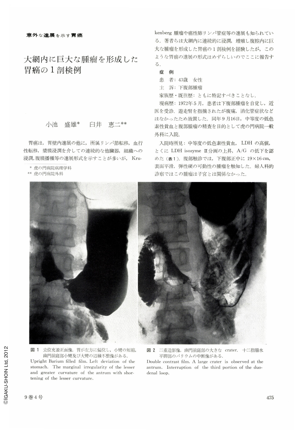
胃癌は,胃壁内進展の他に,所属リンパ節転移,血行性転移,漿膜浸潤を介しての連続的な他臓器,組織への浸潤,腹膜播種等の進展形式を示すことが多いが,Krukenberg腫瘤や癌性肺リンパ管症等の進展も知られている.著者らは大網内に連続的に浸潤,増殖し腹腔内に巨大な腫瘤を形成した胃癌の1剖検例を経験したが,このような胃癌の進展の形式はめずらしいのでここに報告する.
A 43-year-old female complained of a palpable lower abdominal mass in May, 1972, which was diagnosed as gastric carcinoma by means of x-ray examination of the stomach and gastrofiberscopy. The tumor rapidly increased in size to occupy the entire abdominal cavity in Novenmber and she died on Nov. 30, 1972.
Autopsy revealed a gastric carcinoma, located over the entire circumference of the gastric antrum, which showed massive direct invasion into the major omentum measuring 27×21×12 cm in size. On the cut surface the tumor mass in the major omentum showed almost entirely hemorrhagic necrosis. No continuous invasion into other organs except for the transverse colon was noticed.
Histologically, the gastric carcinoma was well-differentiated tubular adenocarcinoma, partly with prominent papillary proliferation and hemorrhagic necrosis in the omental tumor mass.
Several, up to fist-sized metastatic nodes were seen in the liver, where they were medullary and also showed hemorrhagic necrosis. One of the lymph node metastases showed signet-ring cell carcinoma. No peritoneal dissemination was seen.
There was no definite evidence of choriocarcinoma so far as was examined.
Copyright © 1974, Igaku-Shoin Ltd. All rights reserved.