Japanese
English
- 有料閲覧
- Abstract 文献概要
- 1ページ目 Look Inside
I.緒言
脳機能が正常にいとなまれるためには,脳組織の需要に応じてgas交換を行ないうる脳血流が適正に維持される必要がある。そのために正常脳では脳代謝が亢進すればCO2蓄積がおこつて脳血流の増大をきたし,血圧低下にさいしては血管腔が拡大して血流を維持しようとするなど,いわゆる脳血管のautoregulationが有効に作動している。しかし脳の器質的病変,特に脳血管障害では灌流領域にいちじるしい血流動態の変動をきたし,もはやautoregulationは廃絶され,したがつてirrever—sibleな変化を脳組織にあたえると考えられる。このさい脳循環動態に変動をきたす病巣範囲は,多くの場合ある限定された部位である。しかし従来報告されてしいる脳血管障害における脳循環動態の研究は,ほとんどがN2O法による全脳的脳血流量測定法によつてなされたものである。N2O法は正確に脳組織を灌流する血流量を知る有用な測定法ではあるが,得られた値は全脳的なものであつて,必ずしも脳の局所の血流動態を反映したものではない。
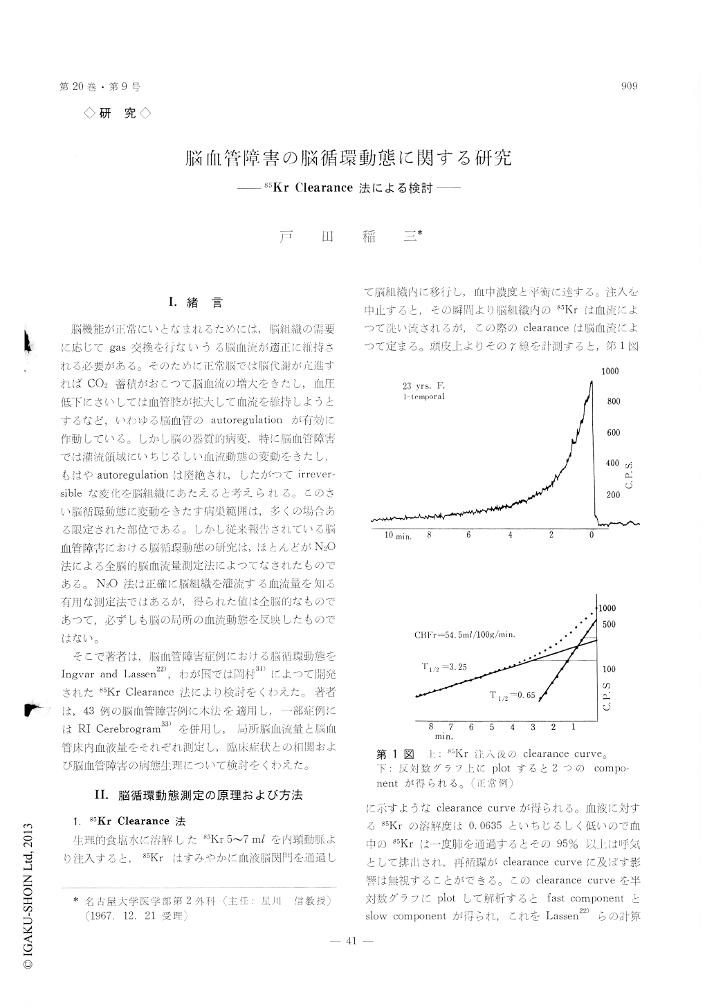
そこで著者は,脳血管障害症例における脳循環動態をIngvar and Lassen22),わが国では岡村31)によつて開発された85Kr Clearance法により検討をくわえた。著者は,43例の脳血管障害例に本法を適用し,一部症例にはRI Cerebrogram33)を併用し,局所脳血流量と脳血管床内血液量をそれぞれ測定し,臨床症状との相関および脳血管障害の病態生理について検討をくわえた。
Regional cerebral blood flow was determined by the method of Lassen and Ingvar in 43 patients with cerebrovascular disease.
The normal blood flow obtained was 54.6 ml/100gm/min in the frontal region and 57.0 ml/100gm/min in the temporal region, respectively.
1) A-V malformation
By means of two techniques, Kr 85 clearance tech-nique by Lassen and Ingvar and nondiffusible RI injection technique by Oldendorf et al, cerebral blood flow was measured in 10 patients with A-V malformation. The former shows the "effective blood flow" which traverses capillaries and involves in the exchange of nutrients and metabolites, the latter shows the blood flow which passes through the intracranial vascular bed and does not move across the blood-brain-barrier.
In all of 10 patients, the dilution curve of RISA bolus obtained from the lesion showed a marked rise in count rate compared with that from the con-tralateral hemisphere, indicating the increased bloodflow through the vascular bed. On the other hand, the curve obtained from Kr 85 clearance technique was found to consists of two components, a large peak, followed by a slower falling phase.
In A-V malformation, the "effective blood flow" decreased due to the blood deprivation by arterio-venous shunt, while the blood flow increased in the vascular bed.
2) Ruptured aneurysm
In 6 patients with ruptued aneurysm, the blood flow decreased remarkably. The mean value of the blood flow was 34.3 ml/100gm/min. In 4 cases of ruptured aneurysm of the anterior communicating artery, bilateral blood flow decreased possibly due to vasospasm of the arteries. It seems to exist a correlation between the level of consciousness and the blood flow. The deeper unconsciousness is acc-ompanied with the lower blood flow. The blood flow was 36.7 ml/100gm/min in the lethalgic state, while 29.6 ml/100gm/min in the comatose state.
3) Obstruction of the internal cartid artery
In 6 patients with obstruction of the internal carotid artery, the blood flow was lower in the lesion side than in the contralateral side. The mean blood flow 29.3 ml/100gm/min on the lesion side, while it was 39.2 ml/100gm/min on the other side.
4) Obstruction of the middle cerebral artery
In all of 8 cases, the blood flow decreased in both hemispheres, especially in the lesion side. The blood flow was 31.6 ml/100mg/min in the lesion side, while it was 42.5 ml/100gm/min in the other side.
5) Cerebral infarction
In 6 patients having hemiplegia following the onset of apoplectic attack without the arteriographic findings of obstruction of arteries, the mean blood flow decreased in both hemispheres. Local decrease of the blood flow within the region corresponding to the clinical symptoms was detected. The blood flow was 40.9 ml/100gm/min in the lesion side and 42.7 ml/100gm/min in the other side.
6) Cerebral arteriosclerosis
In 7 patients with cerebral arteriosclerosis, the mean blood flow was 37.8 ml/100gm/min.

Copyright © 1968, Igaku-Shoin Ltd. All rights reserved.


