Japanese
English
- 有料閲覧
- Abstract 文献概要
- 1ページ目 Look Inside
I.はじめに
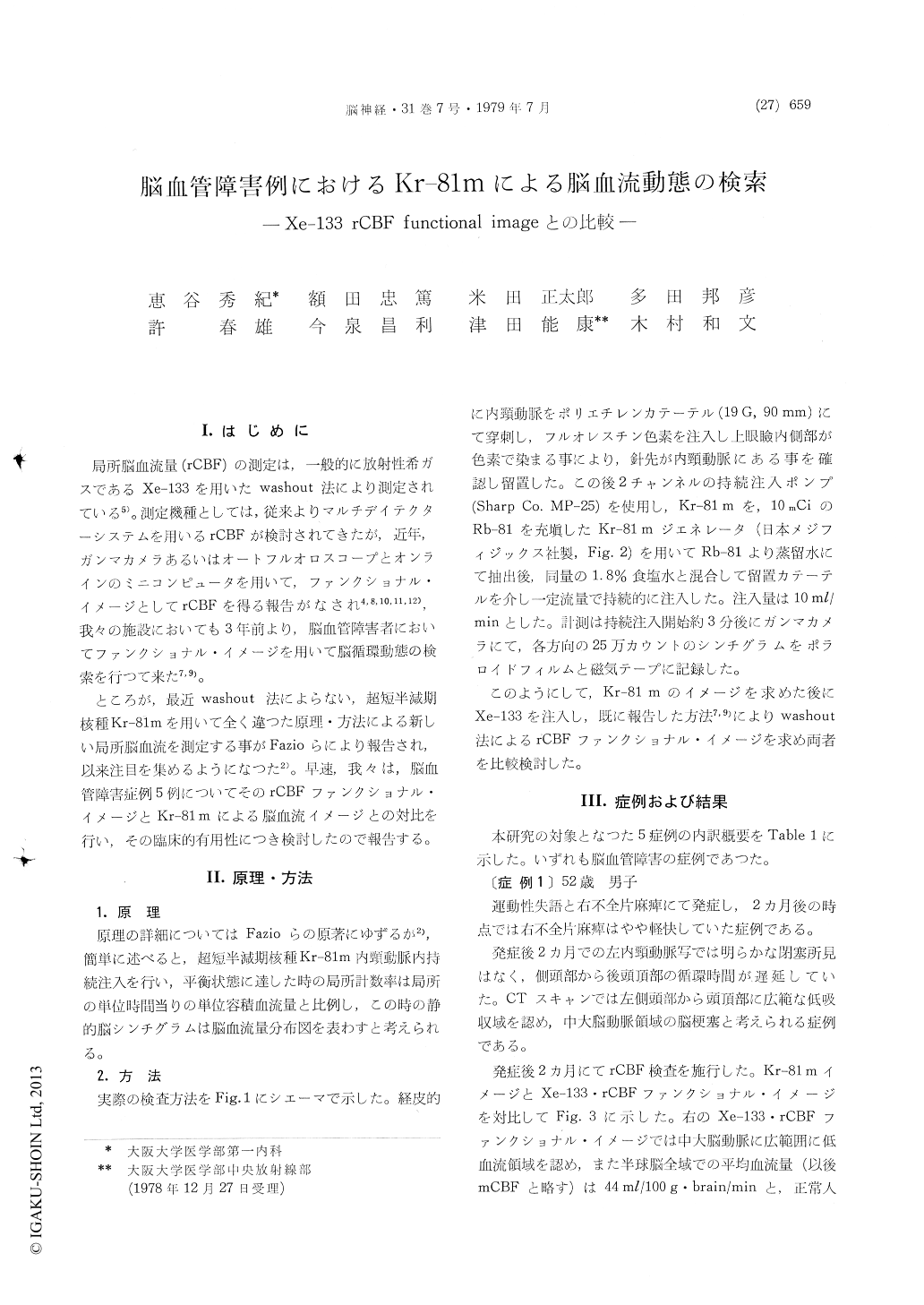
局所脳血流嚢(rCBF)の測定は,一般的に放射性希ガスであるXe−133を用いたwashout法により測定されている5)。測定機種としては,従来よりマルチディテクターシステムを用いるrCBFが検討されてきたが,近年,ガンマカメラあるいはオートフルオロスコープとオンラインのミニコンピュータを用いて,ファンクショナル・イメージとしてrCBFを得る報告がなされ4,8,10,11,12),我々の施設においても3年前より,脳血管障害者においてファンクショナル・イメージを用いて脳循環動態の検索を行つて来た7,9)。
ところが,最近washout法によらない,超短半減期核種Kr−81mを用いて全く違つた原理・方法による新しい局所脳血流を測定する事がFazioらにより報告され,以来注目を集めるようになつた2)。早速,我々は,脳血管障害症例5例についてそのrCBFファンクショナル・イメ—ジとKr−81mによる脳血流イメージとの対比を行い,その臨床的有用性につき検討したので報告する。
Kr-81m brain perfusion image devised by Fazio et al was compared with Xe-133 rCBF functional image in cases with cerebrovascular disease. The Kr-81m eluted by distilled water from Kr-81m generator (Nihon Mediphysics Co.) was mixed by use of Y tube with constant flow, in equal volume of 1.8% NaC1 solution in order to obtain a con-tinuous flow of Kr-81m dissolved in 0.9% Nacl. This Kr-81m in saline was infused by a constant-volume infusion pump (Sharp Co. MP-25) through a small catheter into the internal carotid artery. The flow rate of distilled water for elution of Kr-81m was 10 ml/min. After 3 minutes of the be-ginning of Kr-81m infusion, Kr-81m brain image was obtained by simply collecting counts, up to 250,000 with a gamma camera and recorded on Poraloid film and magnetic tape. After the in-fusion pump was stopped, the radioactivity decreased to the level of the background within 2 minutes. The reproducibility of Kr-81m brain image was excellent in all cases. After Kr-81m brain images were obtained, 5mCi of Xe-133 in saline was in-jected into the catheter and the Xe-l33 rCBF functional images were obtained by processing the data of Xe-133 dynamic images measured by a gamma camera with an on-line mini-computer system.
Four cases with cerebral infarction and one case with transient focal cerebral ischemic attack (TIA) were examined in this study. In the case with TIA (Case 5), Xe-133 rCBF functional image showed no focal low flow area and mean rCBF value was 51 m1/100 g/min. In this case, Kr-81m brain imagealso showed no focal area of reduced activity. In the case with cerebral infarction of MCA territory (Case 1), Kr-81m brain image showed a sharply defined area of reduced activity in the territory of MCA. The rCBF functional image of this case (Case 1) also showed low flow area sharply in the same territory. Similar results were archieved in other cases with cerebral infarction (Case 2, 3 and 4).
In conclusion, Kr-81m brain image well coincided with Xe-133 rCBF functional image in all cases with cerebrovascular disease. Similar to Xe-133 rCBF functional image, Kr-81m brain image was reliable method for detecting the focal hemodvnamicdisorders in cerebrovascular disease. Although this method provided no absolute value of rCBF, the distribution of radioactivity linealy correlated to the rCBF value. This method was simple in the procedure and no computer analysis was required. Due to the very short half life of Kr-81m, the procedure was able to repeat and able to get brain image in multiple views. The reproducibility of Kr-81m brain image was excellent in all cases. A practical disadvantage was that the half life of the parent nuclide (Rb-81) was relatively short (4.6hr.). So Kr-81m generator cannot be used at the area distant from the site of production with a cyclotron.

Copyright © 1979, Igaku-Shoin Ltd. All rights reserved.


