Japanese
English
- 有料閲覧
- Abstract 文献概要
- 1ページ目 Look Inside
I.緒言
最近の脳血管撮影の進歩,普及につれて,頭蓋内の血管性疾患に関する認識が深まり,脳神経外科領域におけるそれら疾患の占める割合は急激に増加している。特にクモ膜下出血の70〜80%を占める脳動脈瘤は,開頭の上直接侵襲を加えることがしだいに常識化されるようになつている。しかしその多くはウィリス動脈輪の前部すなわち天幕上のものであり,天幕下特に脳底動脈より発した動脈瘤については海外の文献もわが国の文献も少ない。今回われわれは脳底動脈動脈瘤の2例を経験したが,内1例に直接手術を試み治癒せしめえたので報告し,私見を述べ,文献的考察を加える。
Two cases of the ruptured aneurysms of the basilar artery were reported. One of which was treated successfully with direct intracranial operation.
Case 1. A 52-year-old man with left hemiplegia and right ocular palsy. He had twice episodes of subarachnoid hemorrhage during these two weeks.
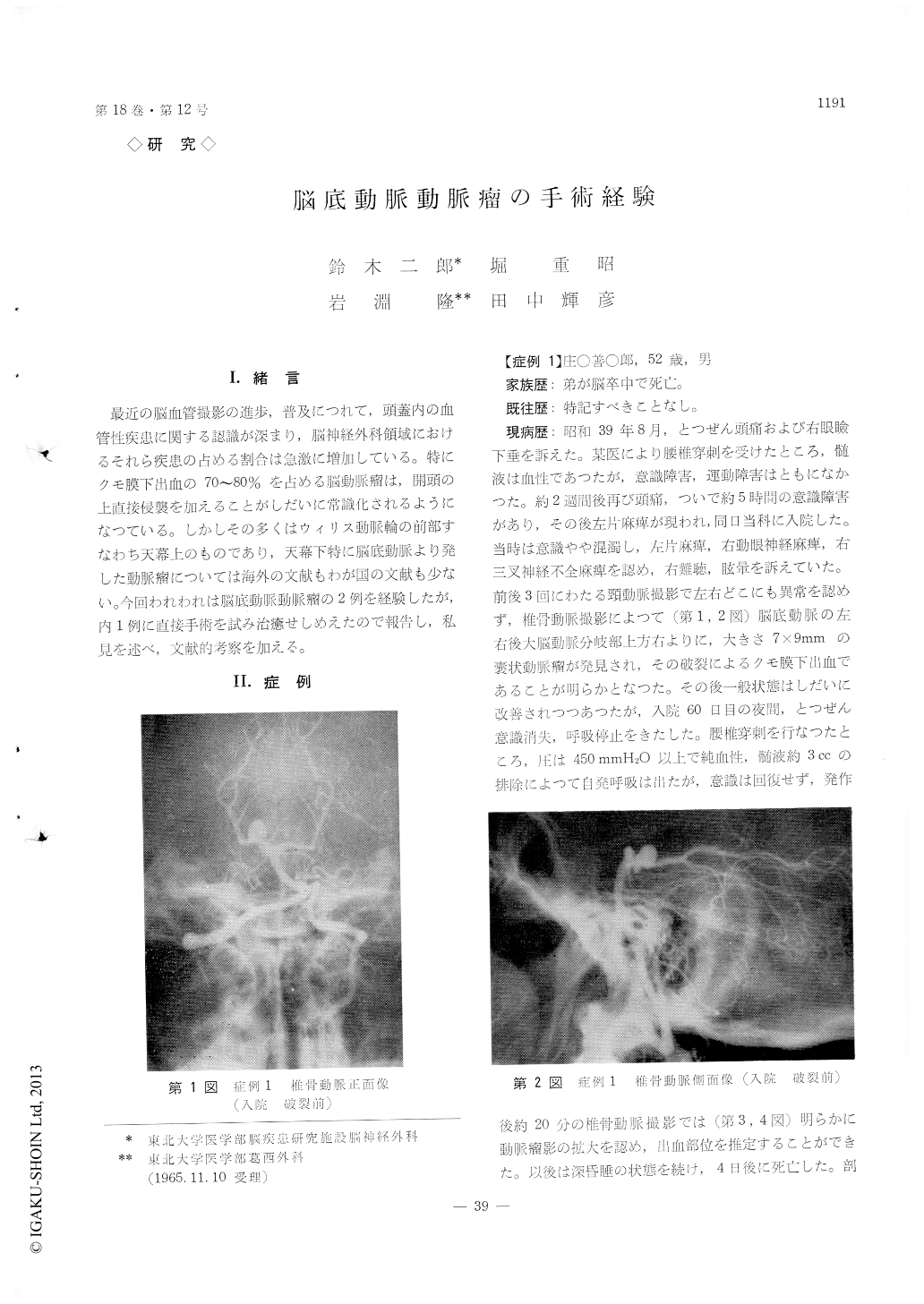
Any aneurysm was not found in the carotid angio-grams but a berry aneurysm (7mm×9mm) was demonstrated at the right upper stem of basilar artery in the vertebral angiography. Unfortunately, he died 4 days after his third attack.
Case 2. A 50-year-old woman. She complained of severe headache and unconciousness 4 hours before admission. Cerebrospinal fluid was hemorrhagic.
A saccular aneurysm (4mm×8mm) arising from basilar artery between right posterior cerebral artery and right superior cerebellar artery was found in ver-tebral angiography.
The direct intracranial operation was performed under hyporthermic anesthesia of 26℃, "Temporal keel form incision" was adopted as the approach to the aneurysm, left temporal love was taken aside by the cerebral spatula, then the basilar artery and the neck of the aneurysm could be seen in the operative field without the injury of tentorial notch.
The neck of aneurysm was clipped successfuly.
She discharged 50 days after operation without any complaint.

Copyright © 1966, Igaku-Shoin Ltd. All rights reserved.


