Japanese
English
- 有料閲覧
- Abstract 文献概要
- 1ページ目 Look Inside
I.はじめに
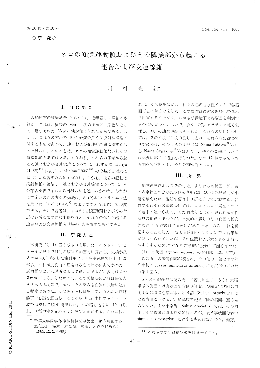
大脳皮質の線維結合については,近年著しく詳細にされた。これは,従来のMarchi法のほかに,染色法として一層すぐれたNauta法が加えられたからである。しかし,これらの方法を用いた研究の多くは投射神経路に関するものであつて,連合および交連神経路に関するものではない。このことは,ネコの知覚運動領ないしその隣接部にもあてはまる。すなわち,これらの領域から起こる連合および交連線維については,わずかにKariya(193611))およびUchishima (1936)25)のMarchi標本に基づいた報告をみるにすぎない。しかも,彼らの記載は投射線維に終始し,連合および交連線維については,その存否を表で示した以外はなにも述べなかつた。したがつてネコのこの方面の知識は,わずかにストリキニン法を用いたGarol (1942)8)によつて支えられている程度である。そこで著者は,ネコの知覚運動預およびその付近の各所に限局的な小傷を与え,それらの傷から起こる連合および交連線維をNauta染色標本で調べてみた。
A series of cat brains with lesions localized sharp-ly to various parts of the sensorimotor cortex and adjacent regions (Gyrus proreus, Gyri sigmoidei anterior et posterior, Gyrus coronalis) were prepared by the Nauta staining method for studying the degenerated association and commissural fibers. The results of the present study are summarized as follows :
(1) The vast majority of association fibers arising from the above-mentioned areas contribute to connec-tion of the same gyrus as lesions and neighbouring gyri. By these fibers, the lateral part of the anterior sigmoid gyrus and the whole extent of the posterior sigmoid and the coronary gyri, namely the leg, arm and face subdivisions of the sensorimotor cortex, are extensively connected to each other. Among these, some fibers extent to the anterior suprasylvian gyrus and the dorsal portion of the anterior ectosylvian gyrus, so that the activity of the sensorimotor cortex may be transmitted to the anterior border of the cortical association and the auditory areas which are located in vicinity.
(2) A relatively small amount of degenerated fibers are traced in such a way as to arise from the coronary gyrus to the anterior half of both walls of the middle suprasylvian sulcus. Considering its course, this fiber group seems to be the superior longitudinal fasciculus described in man and monkey. It is possible to say that fibers from the ventral portion of the coronary gyrus take a longer course backwards along the suclus than ones from the dorsal portion.
(3) No direct fibers from the sensorimotor cortex to the visual area were recognized.
(4) Among association fibers a scanty amount appears terminated in I and II layers. But a large amount seems to make fibers of passage in V and VI layers and preterminal endings in III and IV layers.
(5) It is doubtful of the existence of the commissu-ral fibers from the gyrus proreus and the medial part of both sigmoid gyri. Though fibers from the lateral part of both sigmoid gyri are a few in amount, they connect an area of the contralateral hemisphere corresponding to the site of the lesion. It is noticeable that a relatively large amount from the coronary gyrus were traced in wider area than the extent of the homotopical lesion. Concerning the terminal layers except I and II, they were appro-ximately similar to that of the association fibers.

Copyright © 1966, Igaku-Shoin Ltd. All rights reserved.


