Japanese
English
- 有料閲覧
- Abstract 文献概要
- 1ページ目 Look Inside
I.はじめに
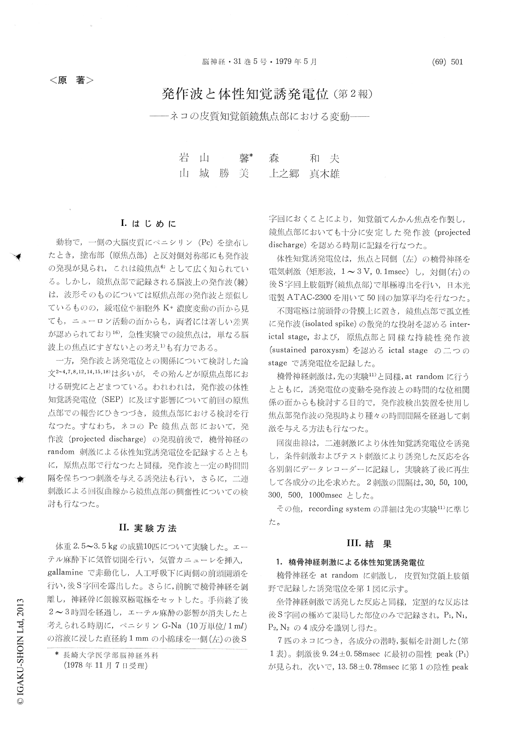
動物で,一側の大脳皮質にペニシリン(Pc)を塗布したとき,塗布部(原焦点部)と反対側対称部にも発作波の発現が見られ,これは鏡焦点6)として広く知られている。しかし,鏡焦点部で記録される脳波上の発作波(棘)は,波形そのものについては原焦点部の発作波と類似しているものの,緩電位や細胞外K+濃度変動の面から見ても,ニューロン活動の面からも,両者には著しい差異が認められており16),急性実験での鏡焦点は,単なる脳波上の焦点にすぎないとの考え1)も有力である。
一方,発作波と書誘発電位との関係について検討した論文2〜4,7,8,12,14,15,18)は多いが,その殆んどが原焦点部における研究にとどまつている。われわれは,発作波の体性知覚誘発電位(SEP)に及ぼす影響について前回の原焦点部での報告にひきつづき,鏡焦点部における検討を行なつた。すなわち,ネコのPc鏡焦点部において,発作波(projected discharge)の発現在前後で,橈骨紳経のrandom刺激による体性知覚誘発電位を記録するとともに,原焦点部で行なったと同様,発作波と一定の時間間隔を保ちつつ刺激を与える誘発法も行い,さらに,二連刺激による回復曲線から鏡焦点部の興奮性についての検討も行なつた。
Epileptogenic foci were induced on the posterior sigmoid gyrus in cats by topical application of penicillin-G-Na (Pc). Primary somatosensory evoked potentials (SEPs) and cortical excitability were studied in the contralateral homologous region, in order to clarify the pathophysiological process underlying"mirror focus".
Primary somatosensory evoked potentials were elicited by electrical stimulation applied to the radial nerve with arbitrary periods following an appearance of spikes in ECoG, and a specific spike-detector was designed for this purpose.
1) Primary somatosensory evoked potentials in control cats were composed of four components, having peak latency of 9.24±0.58 msec (P1), 13.58± 0.78 msec (N1), 19.20±2.82 msec (P2), 33.16±2.64msec (N2) and peak-to-peak amplitude of 33.57± 18.80μV, 130.23±31.40μV, 157.50 ±21.33μV, 80.57 ± 27.00μV, respectively.
2) SEPs elicited during interictal stage where projected ECoG spikes appeared sporadically at the mirror focus, were similar to those elicited before Pc application, in wave form and peak latencies. But an amplitude of all components of SEPs were decreased to 70~80% after appearance of projected ECoG paroxysms.
It was reported that there existed inhibitory mechanisms preventing to a development of epi-leptogenic process in the mirror focus. A decrease in number and synchronization of firing neurons to coming sensory input might result in a diminished amplitude of SEPs.
3) In contrast with the results or original focus, no remarkable change was recognized in SEPs in which radial nerve stimulus was applied immediately after an occurrence of isolated ECoG paroxysms. SEPs obtained after 50 msec or more intervals also revealed similar wave form as compared with those evoked at random intervals.
4) Cortical excitability was measured by recovery curves which were constructed by using measure-ments of SEPs elicited by paired stimuli to radial nerve.
The recovery curves were elevated in all com-ponents of SEPs at the mirror focus during interital stage.
Diencephalon and reticular formation are thought to be important in determining the characteristics of cortical excitability curves, and it was speculated that propagated epileptogenic process to these structures might alter cortical excitabilities via the ascending reticular system or the thalamo-cortical reverberating circuit.
5) Similar to the original focus SEPs disappeared in the mirror focus during tonic-clonic sustained ECoG paroxysms acompanied by surface negative steady potential shifts.

Copyright © 1979, Igaku-Shoin Ltd. All rights reserved.


