Japanese
English
- 有料閲覧
- Abstract 文献概要
- 1ページ目 Look Inside
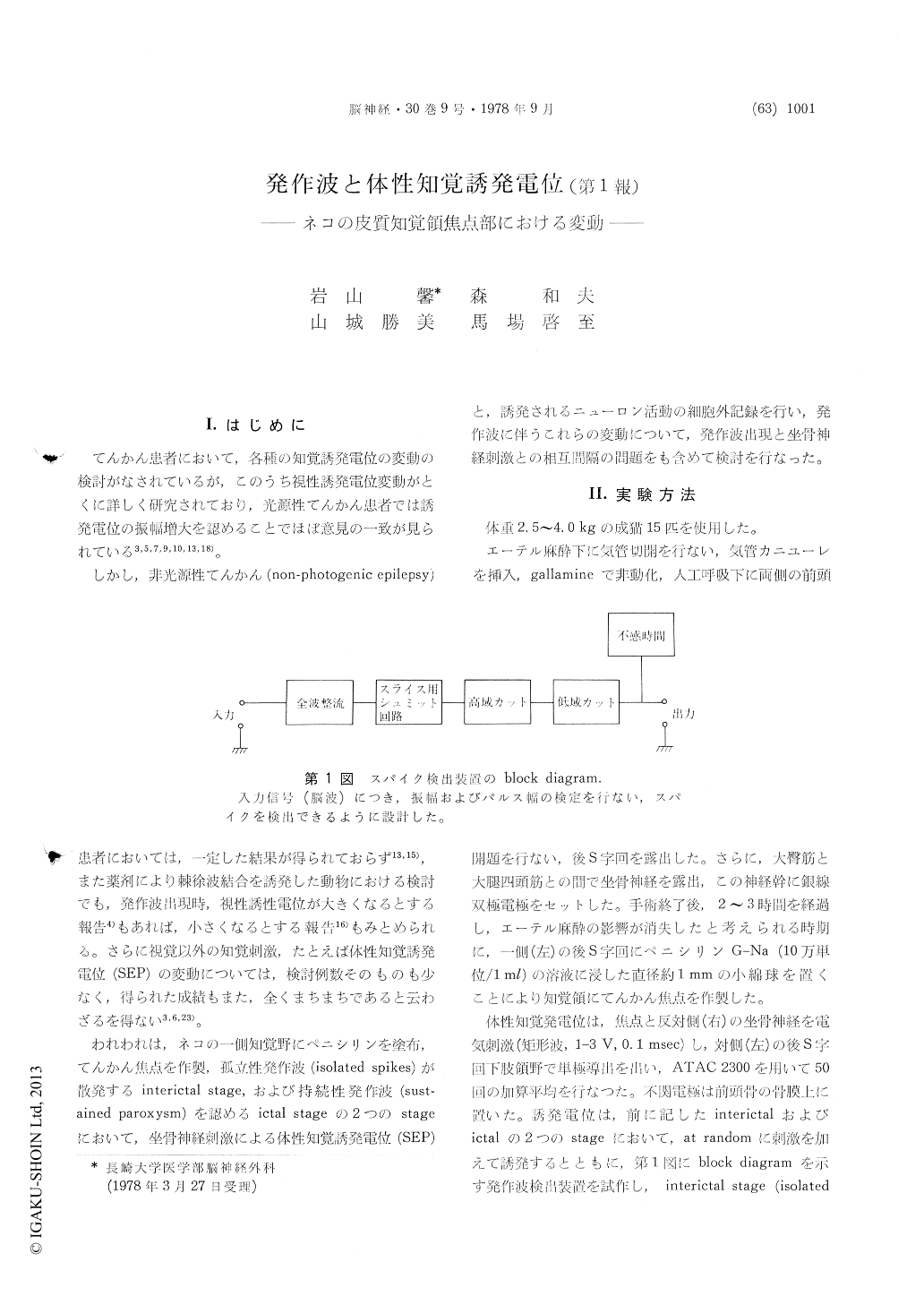
I.はじめに
てんかん患者において,各種の知覚誘発電位の変動の検討がなされているが,このうち視性誘発電位変動がとくに詳しく研究されており,光源性てんかん患者では誘発電位の振幅増大を認めることでほぼ意見の一致が見られている3,5,7,9,10,13,18)。
しかし,非光源性てんかん(non-photogenic epilepsy)患者においては,一定した結果が得られておらず13,15),また薬剤により棘徐波結合を誘発した動物における検討でも,発作波出現時,視性誘性電位が大きくなるとする報告4)もあれば,小さくなるとする報告16)もみとめられる。さらに視覚以外の知覚刺激,たとえば体性知覚誘発電位(SEP)の変動については,検討例数そのものも少なく,得られた成績もまた,全くまちまちであると云わざるを得ない3,6,23)。
An attempt was made to clarify the mutual in-fluences between the epileptic ECoG discharges and the evoked cortical responses. For this purpose, primary somatosensory evoked potentials (SEP) and evoked neuronal activities were recorded simultane-ously on Penicillin-induced focus of the posterior sigmoid gyrus in cats.
1. Primary somatosensory evoked potentials, evo-ked by the contralateral sciatic nerve stimulation,consisted of positive-negative-positive sequence having peak latency of 11.9±0.1 msec (P1), 15.4±0.2 msec (N1), 19.8±0.2msec (P2), followed by another two negative deflections of 24.1±0.2 msec (N2) and 40~50 msec (N3) respectively.
2. In control (prior to application of Pc), extra-cellularlly recorded evoked neurons in the sensory cortex fired during period of 9~40 msec, after con-tralateral sciatic stimulation and the stage of maxi-mal firing was corresponded with the phase of P1 and ascending limb of N1 of the SEP.
3. During interictal stage where isolated ECoG spikes with negative steady potential shifts appeared sporadically, SEP elicited by random stimulus-intervals showed prolongation of latency and in-crease in amplitude of P1 and disappearance of N1. Then large amplitude negative peak of about 500 μV was observed in SEP instead of N2 and N3. In an occasion that sciatic stimulus was applied immediately after the occurrence of isolated ECoG paroxysms, no component of SEP was recognized, but if stimulus was applied about 50 msec after the ECoG paroxysms, SEP in small amplitude was ap-peared. Evoked potentials obtained after 500msec or more revealed similar wave form as compared to those evoked at random intervals.
4. Unitary behavior which was provoked by the stimulation of contralateral sciatic nerve and re-corded with different delay after observing isolated spikes in ECoG, could be differentiated as following three types (Fig.8): Type A unit showed high frequency bursts which occurred about 9msec after stimulation and lasted for 80~90 msec. This type of unit was evoked if the stimulus was applied to 80~90msec after ECoG paroxysm. Type B unit showed maximum firing at about 11 msec, followed by firing with decremental fashion for up to 100 msec. Type C unit showed no evoked neuronal activities. All of the evoked neuronal activities recorded immediately after ECoG paroxysms showed type C firing. The more the cell showing type A firing increased, the longer the intervals between ECoG paroxysms and stimulation was used. It was considered that the high amplitude negative poten-tials in SEP were represented epileptiform ECoG discharge itself, because in accordance with this stage, high frequency epileptic neuronal burst was also identified in the present study.
5. It was also noted that SEP disappeared during tonic-clonic sustained ECoG paroxysms acompanied by surface negative steady potential shifts.
Copyright © 1978, Igaku-Shoin Ltd. All rights reserved.