Japanese
English
- 有料閲覧
- Abstract 文献概要
- 1ページ目 Look Inside
I.はじめに
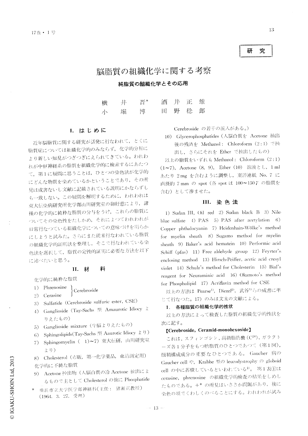
近年脳脂質に関する研究が活発に行なわれて,とくに脂質症については組織化学的のみならず,化学的分析により新しい知見がつぎつぎにえられてきている。われわれが中枢神経系の脂質を組織化学的に検索するにあたつて,第1に疑問に思うことは,ひとつの染色法が化学的にどんな物質を染めているかということであり,その所見は成書ないし文献に記載されている説明にかならずしも一致しない。この疑問を解明するために,われわれは東大伝染病研究所化学部山川研究室の御好意により,諸種の化学的に純粋な脂質の分与をうけ,これらの脂質についてその染色性をたしかめ,それによつてわれわれが日常行なつている組織化学についての意味づけを明らかにしようと試みた。さらにまた従来行なわれている脂質の組織化学的証明法を整理し,そこで行なわれている染色法を選択して,脂質の定性的証明に必要な方法を以下に述べたいと思う。
Histochemistry of lipid is important for the patho-logical study of the brain disease, especially of lipidosis and demyelinating disease. There are numbers of histochemical methods for lipid in the book of histo-chemistry, however, the staining properties of chemi-cally pure substances have not been well known.
This communication deals with the histochemistry of various kinds of pure lipids which exist in the central nervous system. The histochemical properties of each lipid are shown in Table 1~7, while Table 8~12 show the reaction of each staining method upon several kinds of lipids.
On the basis of the data above mentioned, and for the purpose of identification of lipid in the brain tissue qualitatively, the procedures which consist of several kinds of staining methods and the treatment of section in some fat solvent, are deviced as follows.
The section has to be frozen.
1) For the identification of phospholipid, Sudan black B which stains phospholipid intensely within 10 minutes, is the most available at the first step. The performic-acid Schiff reaction is applied at the second step, because of its specific, positive reaction upon glycerophosphatides. Lecithine proves its presence by the positive copper phthalocyanin reaction, while sphingomyelin which also reacts positively upon copper phthalocyanin reaction, is certified in the case of negative performic-acid-Schiff reaction or after the treatment of section in hot ether 40℃ for 1 hour. The interference of cholesterin and cholesterin ester can be eliminated by the treatment of section in cold acetone for 1 hour beforehand, because cholesterin and cholesterin ester give the positive reaction by Sudan black B staining.
2) For the examination of glycolipid, the meta-chromasia by means of cresyl violet in 1% acetic acid solution, Hirsch-Peiffer's method, seems to be the most useful for the differentiation between glyco-lipids, because cerebroside-sulfuric-ester gives the distinctive brown metachromasia and ganglioside shows the γ-metachromasia by this method. Holländer's acriflavin method for cerebroside-sulfuric-ester is ex-actly specific. As far as the differentiation between glycolipids is concerned, glycerophosphatides which show also γ-metachromasia with this staining, should be extracted from the section beforehand by the treatment of section in hot ether 40℃ for 1~2 hours. Cerebroside reacts negatively upon a lot of lipid staining, except for PAS reaction and Nile blue sulfate staining, therefore the presence of cerebroside in tissue is often difficult to prove and remains only suspicious.

Copyright © 1965, Igaku-Shoin Ltd. All rights reserved.


