Japanese
English
- 有料閲覧
- Abstract 文献概要
- 1ページ目 Look Inside
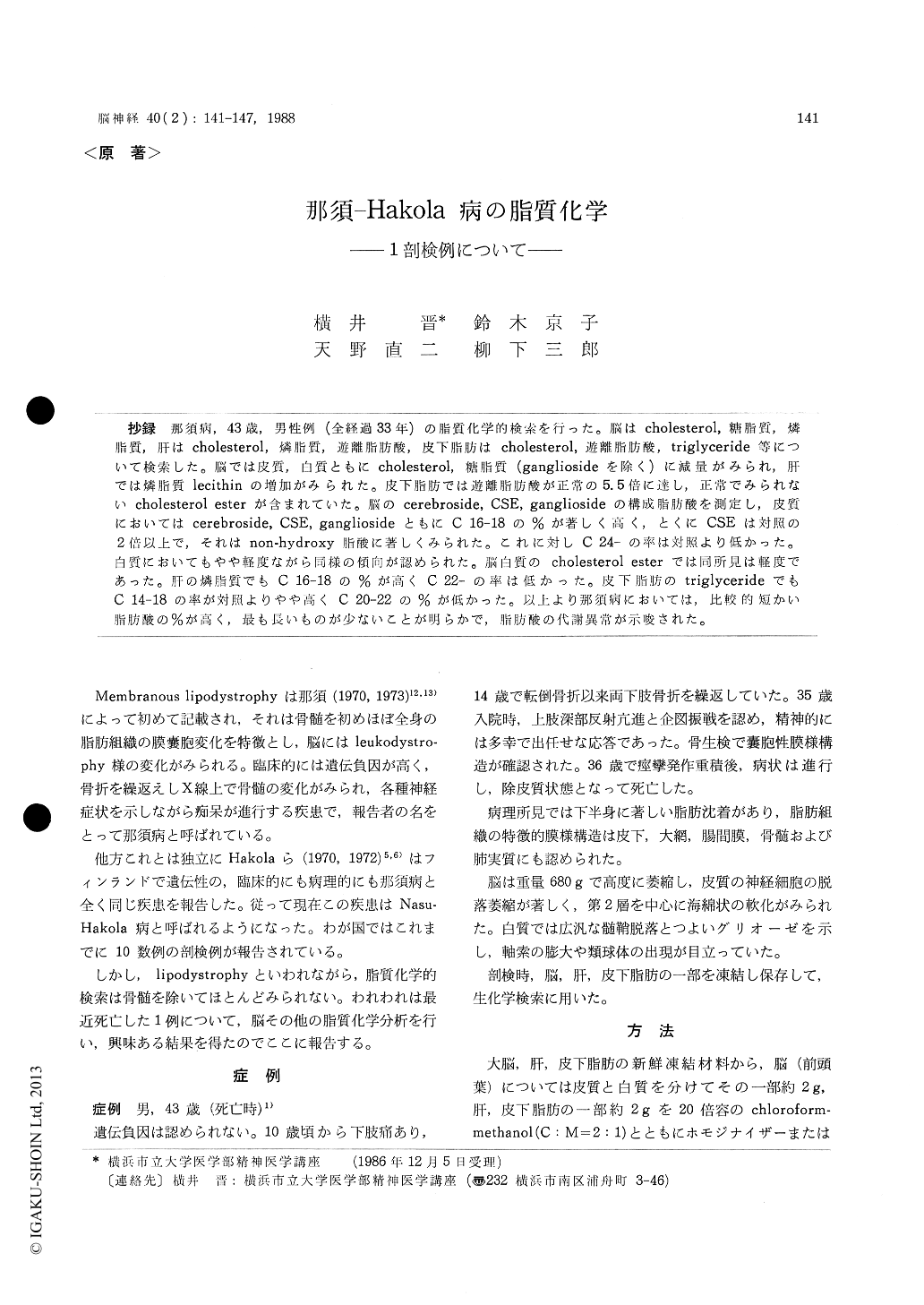
抄録 那須病,43歳,男性例(全経過33年)の脂質化学的検索を行った。脳はcholesterol,糖脂質,燐脂質,肝はcholesterol,燐脂質,遊離脂肪酸,皮下脂肪はcholesterol,遊離脂肪酸,triglyceride等について検索した。脳では皮質,白質ともにcholesterol,糖脂質(gangliosideを除く)に減量がみられ,肝では燐脂質lecithinの増加がみられた。皮下脂肪では遊離脂肪酸が正常の5.5倍に達し,正常でみられないcholesterol esterが含まれていた。脳のcerebroside, CSE, gangliosideの構成脂肪酸を測定し,皮質においてはcerebroside, CSE, gangliosideともにC16-18の%が著しく高く,とくにCSEは対照の2倍以上で,それはnon-hydroxy脂酸に著しくみられた。これに対しC24-の率は対照より低かった。白質においてもやや軽度ながら同様の傾向が認められた。脳白質のcholesterol esterでは同所見は軽度であった。肝の燐脂質でもC16-18の%が高くC22-の率は低かった。皮下脂肪のtriglycerideでもC14-18の率が対照よりやや高くC20-22の%が低かった。以上より那須病においては,比較的短かい脂肪酸の%が高く,最も長いものが少ないことが明らかで,脂肪酸の代謝異常が示唆された。
Lipid chemical analysisof a case of membranous lipodystrophy (Nasu-Hakola disease) was reported. The case is a 43 yr-old man (at death). Onset ofthe disease was at his age of 10 when he had complained of leg pain. Since his age of 14, he had bone fractures of the lower extremities many times. At the age of 35, he was admitted to a hospital for the neurological examination. At that time, he showed exaggerated tendon reflexes and intension tremor of the upper extremities. He was euphoric and demented. His IQ was 31. Histological examination of biopsy specimen from the bone marrow revealed the typical membrano-cystic lesion. He had status epilepticus which was followed by comatous state for a week. The disease progressed gradually and he fell into de-corticated state. He died at the age of 43. The total clinical course was 33 years.
The brain weighed 680 g. Remarkable atrophy was observed in the whole brain. Loss of nerve cells in the upper layers of the cortex was marked. There were loss of myelin sheath and severe gliosis in the white matter.
A part of the brain, liver and subcutaneous fat was frozen at the time of autopsy for the chemi-cal examination.
Qantitative determination of cholesterol, glyco-lipid, phospholipid, free fatty acid in the brain and liver, cholesterol, free fatty acid, triglyceride in the subcutaneous fat were carried out.
Cholesterol was decreased about 33% in the cortex and 42% in the white matter in comparing with those of controls and the content of cerbro-side in the white matter was about 1/3 of that of controls. Ganglioside in the cortex was well preserved. Lecithin was increased about 5 times of control. In the liver, lecithin was increased about 3. 2 times and cephalin was decreased about 1/6 of control. Free fatty acid in the subcutaneous fat was 5.5 times as much as that of control and cholesterolester which was not found in control materials, was seen in the subcutaneous fat of the patient.
Fatty acids of cerebroside, CSE, phospholipid, triglyceride and cholesterolester were analysed by gaschromatography. Percentage of C 16-18 fatty acids of CSE and cerebroside in the cortex was remarkably high, namely 22.54% and 12.09% (control : 7.32% and 9.77%) and the rate of very long-fatty acids C 24- was low, they are 62.71% and 61.03% (control : 74.59% and 70.91%). The same tendency was observed in the white matter where % of CSE was 17.04% (control : 11.08%) and of cerebroside was 11.48% (control : 6.20%). The similar findings were observed in relatively short-chain fatty acid (C 16) of ganglioside. These findings were more marked in the non-hydroxy fatty acids. Fatty acids of cholesterolester had a slight degree of the same tendency. High percent-age of C 16-18 of phospholipid, 81.20% (control : 69.60%) in the liver was marked and the rate of C-22 was low, namely 6.25% (control : 15.10%). Analysis of triglyceride of subcutaneous fat show-ed slightly high % of C 14-18 and low rate ofC 20-22.
Based uppon these findings mentioned above, it was suggested that some metabolic abnormality of lipids exsists in Nasu-Hakola disease.

Copyright © 1988, Igaku-Shoin Ltd. All rights reserved.


