Japanese
English
- 有料閲覧
- Abstract 文献概要
- 1ページ目 Look Inside
末梢神經のレ線像は,(6)氣腦撮影Pneumoencephalo graphyに際して,視神經交叉部槽Cisterna chitmsmatis内に視神經,視神經交叉部及び視神經索像が出現する,又腦脚間槽Cisterna interpeduncularis内に動眼神經像が現れる,又沃度油蜘蛛膜下腔撮影法に際しても,脊髓神經が硬膜穿孔部で撮影されることがある。此等は或る目的の操作中神經が偶然にも撮影されたのである。
末梢神經のレ線寫眞は人體に於て初めて,齋藤,神川及び加藤(1931)に依て撮影された。造影劑は,Lipiodol乳劑L'Ombre(1.2.)(7)使用された。
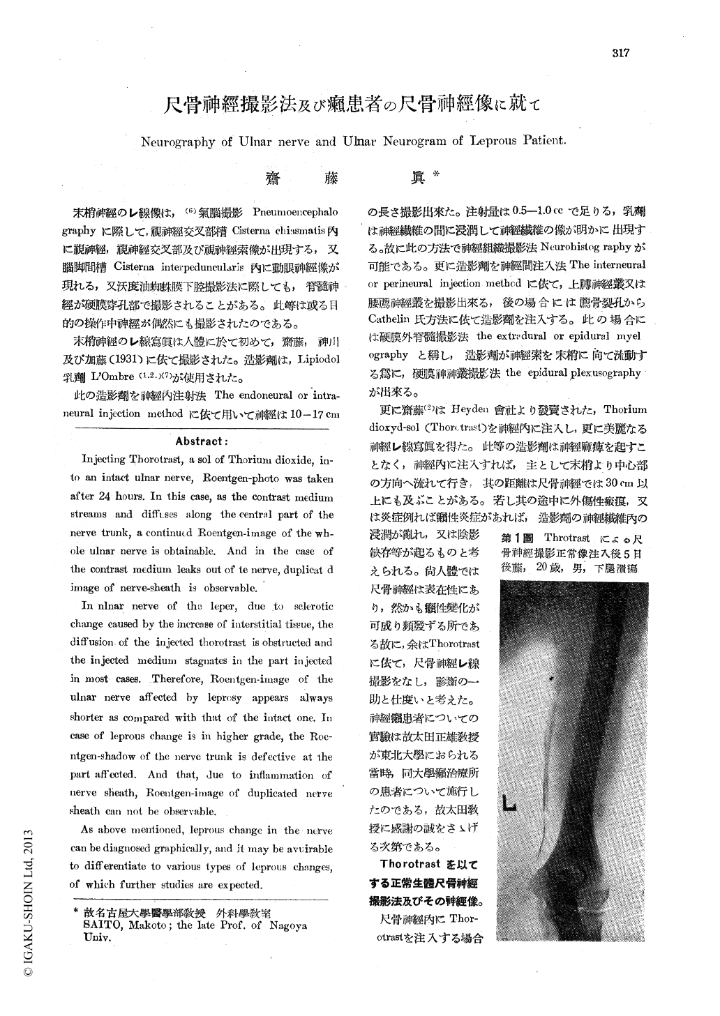
Injecting Thorotrast, a sol of Thorium dioxide, in-to an intact ulnar nerve, Roentgen-photo was taken after 24 hours. In this case, as the contrast medium streams and diffuses along the central part of the nerve trunk, a continued Roentgen-image of the wh-ole ulnar nerve is obtainable. And in the case of the contrast medium leaks out of to nerve, duplicat d image of nerve-sheath is observable.
In nlnar nerve of the leper, due to sclerotic change caused by the increase of interstitial tissue, the diffusion of the injected thorotrast is obstructed and the injected medium stagnates in the part injected in most cases. Therefore, Roentgen-image of the ulnar nerve affected by leprosy appears always shorter as compared with that of the intact one. In case of leprous change is in higher grade, the Roe-ntgen-shadow of the nerve trunk is defective at the part affected. And that, due to inflammation of nerve sheath, Roentgen-image of duplicated nerve sheath can not be observable.
As above mentioned, leprous change in the nerve can be diagnosed graphically, and it may be avuirable to differentiate to various types of leprous changes, of which further studies are expected.

Copyright © 1950, Igaku-Shoin Ltd. All rights reserved.


