Japanese
English
- 有料閲覧
- Abstract 文献概要
- 1ページ目 Look Inside
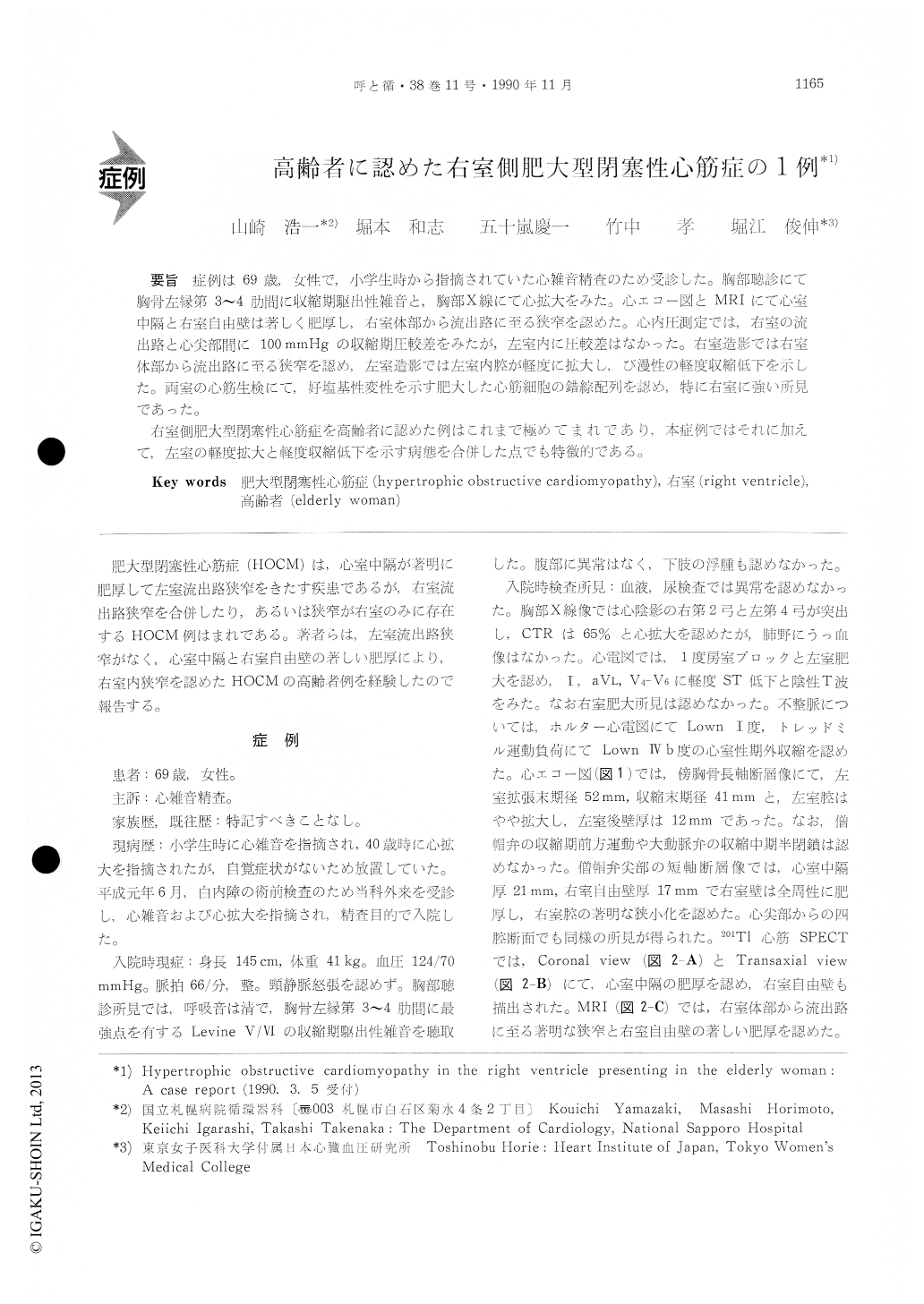
症例は69歳,女性で,小学生時から指摘されていた心雑音精査のため受診した。胸部聴診にて胸骨左縁第3〜4肋問に収縮期駆出性雑音と,胸部X線にて心拡大をみた。心エコー図とMRIにて心室中隔と右室自由壁は著しく肥厚し,右室体部から流出路に至る狭窄を認めた。心内圧測定では,右室の流出路と心尖部間に100mmHgの収縮期圧鮫差をみたが,左室内に圧較差はなかった。右室造影では右室体部から流出路に至る狭窄を認め,左室造影では左室内腕が軽度に拡大し,び漫性の軽度収縮低下を示した。両室の心筋生検にて,好塩基性変性を示す肥大した心筋細胞の錯綜配列を認め,特に右室に強い所見であった。
右室側肥大型閉塞性心筋症を高齢者に認めた例はこれまで極めてまれであり,本症例ではそれに加えて,左室の軽度拡大と軽度収縮低下を示す病態を合併した点でも特徴的である。
A 69-year-old woman was admitted to the hospi-tal for evaluation of a cardiac nturmur, which had been indicated at her childhood.
On physical examination, a systolic ejection mur-mur was heard on the lower left sternal border. Chest roentgenogram revealed a cardiomegaly. Echo-cardiography and MRI showed a thicking of the in-terventricular septum and the right ventricular (RV) free wall, which obstructed the RV body.
At cardiac catheterization, a systolic pressure gra-dient of 100 mmHg was shown between the RV out-flow tract and the apex. There was no pressure gradient in the left ventricle (LV). RV angiocar-diograms disclosed a severe obstruction of the RV body, while LV angiocardiograms showed a slight enlargement of the LV with a diffuse hypokinesis of its wall.
Myocardial biopsy of both ventricles revealed a bizarre hypertrophy of myocytes and disorganization. The histological findings were more conspicuous inthe RV.
Based on these findings, this case was diagnosed as hypertrophic obstructive cardiomyopathy (HOCM) in the RV.
HOCM in the RV presenting in the elderly, is extremely rare. Our case is also characterized by a concomitant LV involvement, which demonstrated the slightly enlarged LV and the diffuse hypokin-esis of its wall.

Copyright © 1990, Igaku-Shoin Ltd. All rights reserved.


