Japanese
English
- 有料閲覧
- Abstract 文献概要
- 1ページ目 Look Inside
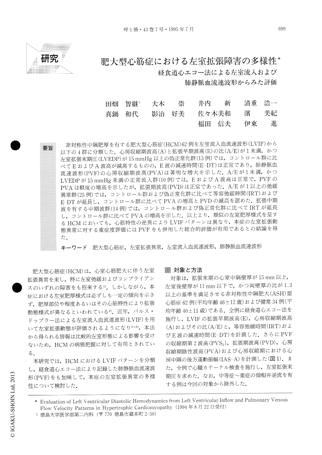
非対称性中隔肥厚を有する肥大型心筋症(HCM)62例を左室流入血流速波形(LVIF)から以下の4群に分類した.心房収縮期波高(A)と拡張早期波高(E)の比(A/E)が1未満,かつ左室拡張末期圧(LVEDP)が15mmHg以上の偽正常化群(13例)では,コントロール群に比べてEおよびA波高が減高するものの,E波の減速時間(E-DT)は正常であり,肺静脈血流速波形(PVF)の心房収縮期波高(PVA)は著明な増大を示した.A/Eが1未満,かつLVEDPが15mmHg未満の正常流入群(10例)では,EおよびA波高は正常で,PVFのPVAは軽度の増高を示したが,拡張期波高(PVD)は正常であった.A/Eが1以上の弛緩異常群(25例)では,コントロール群および偽正常化群に比べて等容弛緩時間(IRT)およびE-DTが延長し,コントロール群に比べてPVAの増高とPVDの減高を認めた.拡張中期波を有する中期波群(14例)では,コントロール群および偽正常化群に比べてIRTが延長し,コントロール群に比べてPVAの増高を示した.以上より,類似の左室肥厚様式を呈するHCMにおいても,心筋特性の差異によりLVIFパターンは異なり,本症の左室拡張動態異常に対する重症度評価にはPVFをも併用した総合的評価が有用であるとの結論を得た.
To evaluate the characteristics of left ventricular diastolic hemodynamics in hypertrophic cardiomyopa-thy (HCM), we recorded left ventricular inflow (LVIF) and pulmonary venous flow (PVF) velocity patterns by transesophageal echocardiography in 62 patients with asymmetric septal hypertrophic type of HCM and 34 normal controls. The patients were divided into four groups according to the LVIF pattern, that is, 1) pseudonormal group; 13 patients with the ratio of an atrial contraction (A) wave to an early diastolic (E) wave (A/E)≦1 and left ventricular enddiastolic pres-sure (LVEDP)≧15mmHg, 2) normal pattern group; 10 patients with the A/E ratio 1 and LVEDP<15mmHg, 3) relaxation failure group; 25 patients with the A/E ratio> 1, and 4) mid-diastolic wave group; 14 patients with mid-diastolic wave. The peak velocity of E wave in pseudonormal, relaxation failure and mid-diastolic wave groups was significantly smaller than that of the control group. Deceleration time of E wave and isovolurnic relaxation time were significantly more prolonged in relaxation failure and mid-diastolic wave groups than in pseudonormal and control groups. The peak velocity of the diastolic wave of PVF in relaxation failure and mid-diastolic wave groups was significantly decreased compared with that of the control group, and was significantly more increased in the pseudonormal group than in the relaxation failure and mid-diastolic wave groups. Compared with the control group, the peak velocity of atrial contraction wave of PVF was significantly increased in all HCM groups, particulary in the pseudonormal group. LVEDP was highest in the pseudonormal group, followed by the mid-diastolic wave, relaxation failure and normal pattern groups, respectively.
In conclusion, combined analysis of LVIF and PVF patterns made it possible to evaluate the various abnor-malities of left ventricular diastolic hemodynamics in HCM.

Copyright © 1995, Igaku-Shoin Ltd. All rights reserved.


