Japanese
English
- 有料閲覧
- Abstract 文献概要
- 1ページ目 Look Inside
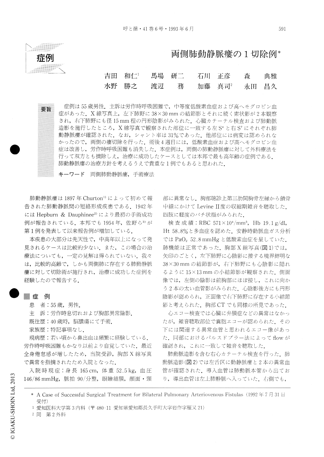
症例は55歳男性.主訴は労作時呼吸困難で,中等度低酸素血症および高ヘモグロビン血症があった.X線写真上,左下肺野に38×30 mmの結節影とそれに続く索状影が2本観察され,右下肺野にも径15mm程の円形陰影がみられた.心臓カテーテル検査および肺動脈造影を施行したところ,X線写真で観察された部位に一致する左S4と右S7にそれぞれ肺動静脈瘻が確認された.なお,シャント率は31%であった.他部位には病変は認められなかったので,両側の瘻切除を行った.術後4週目には,低酸素血症および高ヘモグロビン血症は改善し,労作時呼吸困難も消失した.本症例は,両側の肺動静脈瘻に対して外科療法を行って双方とも摘除しえ,治療に成功したケースとしては本邦で最も高年齢の症例である.肺動静脈瘻の治療方針を考えるうえで貴重な1例でもあると思われた.
A 55-year-old man was admitted to our hospital with chief complaints of exertional dyspnea and chest x-ray abnormalities. On physical examinations, clubbing and systolic murmur were detected. The patient's hemoglo-bin rose to a level of 19.0g/dl, with a rise in hematocrit to 58.8%. While the patient was breathing room air, the PaO2 was 52.8 mmHg, the PaCO2 33.3 mmHg and the pH 7.43. In chest x-ray film, oval well-margined 38×30 mm density with enlarged afferent and efferent vessels in the left upper lobe (lts4) and 15×13 mm “coin lesion” in the right lower lobe (rts7) were observed.

Copyright © 1993, Igaku-Shoin Ltd. All rights reserved.


