Japanese
English
- 有料閲覧
- Abstract 文献概要
- 1ページ目 Look Inside
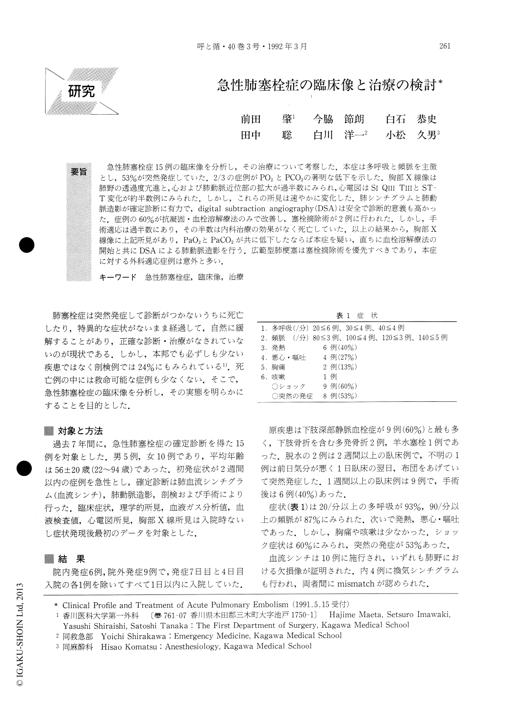
急性肺塞栓症15例の臨床像を分析し,その治療について考察した.本症は多呼吸と頻脈を主徴とし,53%が突然発症していた.2/3の症例がPO2とPCO2の著明な低下を示した.胸部X線像は肺野の透過度亢進と,心および肺動脈近位部の拡大が過半数にみられ,心電図はSI QⅢ TⅢとST—T変化が約半数例にみられた.しかし,これらの所見は速やかに変化した.肺シンチグラムと肺動脈造影が確定診断に有力で,digital subtraction angiography(DSA)は安全で診断的意義も高かった.症例の60%が抗凝固・血栓溶解療法のみで改善し,塞栓摘除術が2例に行われた.しかし,手術適応は過半数にあり,その半数は内科治療の効果がなく死亡していた.以上の結果から,胸部X線像に上記所見があり,PaO2とPaCO2が共に低下したならば本症を疑い,直ちに血栓溶解療法の開始と共にDSAによる肺動脈造影を行う.広範型肺梗塞は塞栓摘除術を優先すべきであり,本症に対する外科適応症例は意外と多い.
The clinical profiles of 15 patients with acute pulmo-nary embolism (APE) were analysed. The most com-mon symptoms of APE were tachypnea and tachycardia with sudden onset. Both P02 and PCO2 had decreased in almost all patients (mean P02: 50mmHg, PCO2: 30mmHg). Chest roentgenogram (X-P) revealed hyper-lucency of the lung field, prominence of proximal pul-monary artery and cardiac enlargement. ECG showed SI QⅢ TⅢ and ST-T changes in half of the cases. These changes, however, disappeared within 4 days in most patients. Lung scan and digital subtraction pulmo-nary angiography were useful for the diagnosis.

Copyright © 1992, Igaku-Shoin Ltd. All rights reserved.


