Japanese
English
- 有料閲覧
- Abstract 文献概要
- 1ページ目 Look Inside
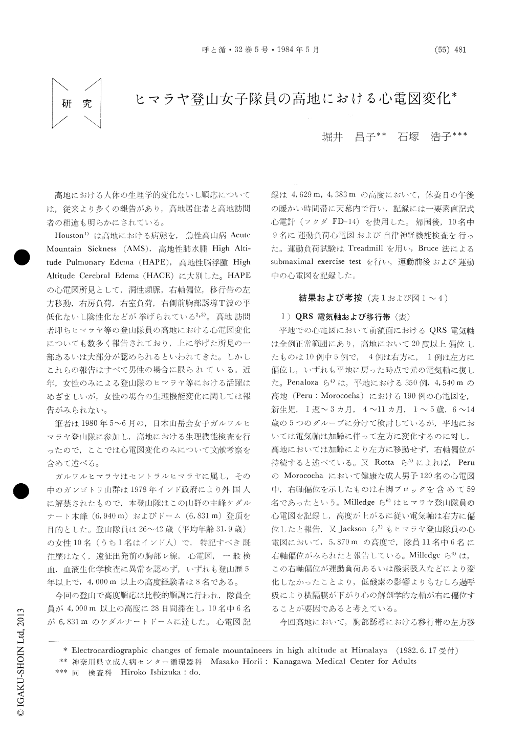
高地における人体の生理学的変化ないし順応については,従来より多くの報告があり,高地居住者と高地訪問者の相違も明らかにされている。
Houston1)は高地における病態を,急性高山病AcuteMountain Sickness(AMS),高地性肺水腫High Alti—tude Pulmonary Edema(HAPE),高地性脳浮腫HighAltitude Cerebral Edcma(HACE)に大別した。HAPEの心電図所見として,洞性頻脈,右軸偏位,移行帯の左方移動,右房負荷,右室負荷,右側前胸部誘導T波の平低化ないし陰性化などが挙げられている2,3)。高地訪問者即ちヒマラヤ等の登山隊員の高地における心電図変化についても数多く報告されており,上に挙げた所見の一部あるいは大部分が認められるといわれてきた。しかしこれらの報告はすべて男性の場合に限られている。近年,女性のみによる登山隊のヒマラヤ等における活躍はめざましいが,女性の場合の生理機能変化に関しては報告がみられない。
Electrocardiograms of the ten female mountain-eers (nine Japanese and one Indian, mean age31.9 years, range 26 to 42 years) were recorded in high altitudes at 4383 meters and 4629 meters above the sea level, respectively.
Right axis deviation in four subjects, a shift of transitional zone to the left in five subjects, and T wave inversion of flattening in the right precor-dial leads were found in five subjects. Further-more, a decrease in the T wave amplitude and biphasic T wave in the left precordial leads in three climbers. Above changes were seen either in isolation or in combination in the same sub-jects. All abnormal changes were normalized upon arrival at sea level, except for one person in which T wave inversion in the right precordial leads persisted four months afterwards. Similar changes have reported in the studies in male mountaineers.
The significance of these changes on electrocar-diograms was discussed. Although the right axis deviation, shift of transitional zone to the left, and T wave inversion or flattening in the right precordial leads are well established observation in patients who suffer from "High Altitude Pufino-nary Edema", our findings secures to have had no relation to "High Altitude Pulmonary Edema" in the light of their physical examinations.
Similar findings among the male mountaineers have also been reported. Therefore, our impression is that there is no essential difference between male mountaineers and female mountaineers.
In the light of experimental investigations performed by Laciga et al in a low atmospheric pressure, we are inclined to believe that respira-tory alkalosis and increase of heart rate both secondary to hypoxia could have accounted for such ECG changes. In addition a change in the sympathetic tone might have also played a role on appearances of the above changes.

Copyright © 1984, Igaku-Shoin Ltd. All rights reserved.


