Japanese
English
- 有料閲覧
- Abstract 文献概要
- 1ページ目 Look Inside
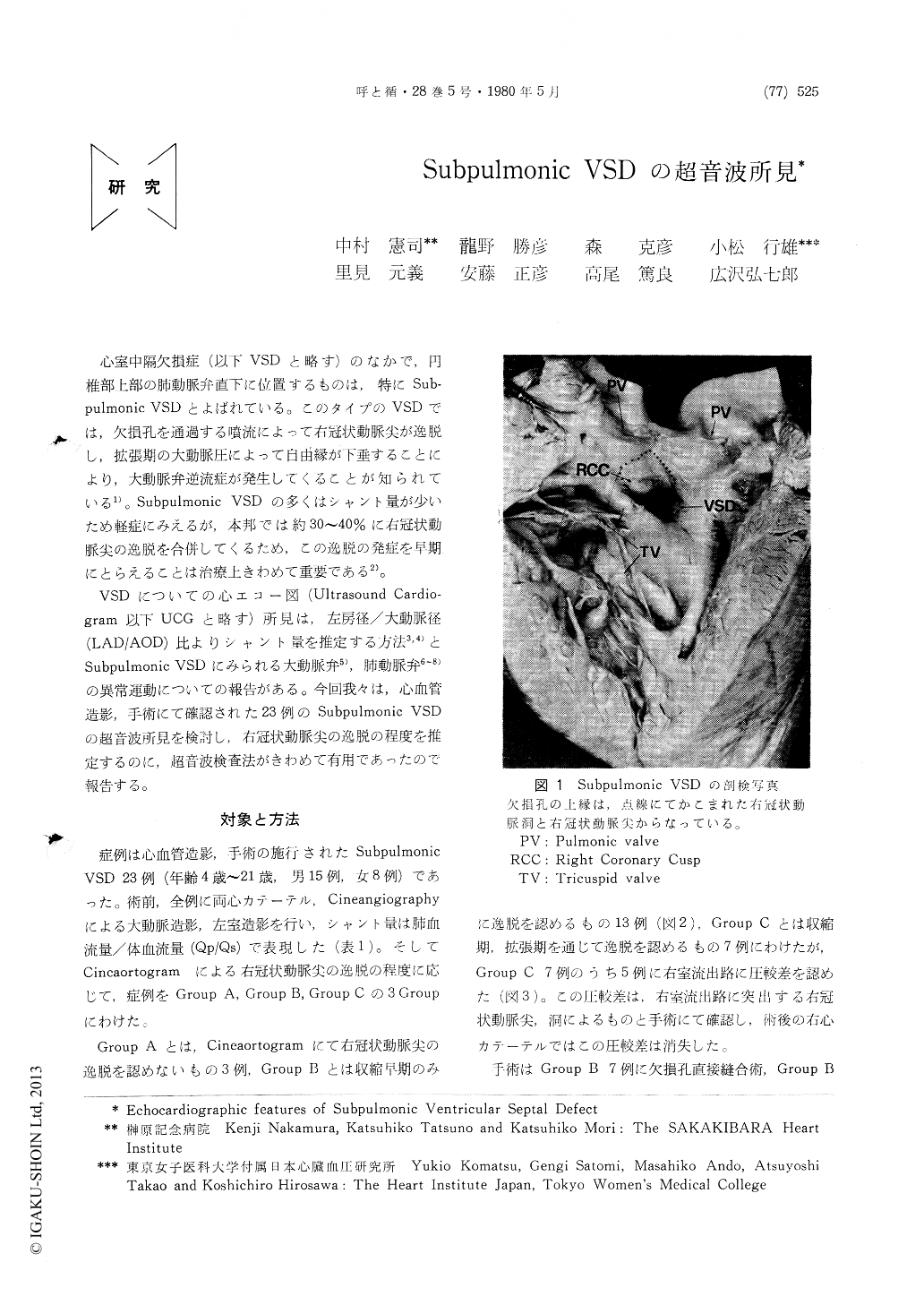
心室中隔欠損症(以下VSDと略す)のなかで,円椎部上部の肺動脈弁直下に位置するものは,特にSub—pulmonic VSDとよばれている。このタイプのVSDでは,欠損孔を通過する噴流によって右冠状動脈尖が逸脱し,拡張期の大動脈圧によって自由縁が下垂することにより,大動脈弁逆流症が発生してくることが知られている1)。Subpulmonic VSDの多くはシャント量が少いため軽症にみえるが,本邦では約30〜40%に右冠状動脈尖の逸脱を合併してくるため,この逸脱の発症を早期にとらえることは治療上きわめて重要である2)。
VSDについての心エコー図(Ultrasound Cardio—gram以下UCGと略す)所見は,左房径/大動脈径(LAD/AOD)比よりシャント量を推定する方法3,4)とSubpulmonic VSDにみられる大動脈弁5),肺動脈弁6〜8)の異常運動についての報告がある。今回我々は,心血管造影,手術にて確認された23例のSubpulmonic VSDの超音波所見を検討し,右冠状動脈尖の逸脱の程度を推定するのに,超音波検査法がきわめて有用であったので報告する。
Echocardiograms in twenty three patients with subpulmonic VSD were analyzed to correlate with the angiographic and hemodynamic findings.
Based on the angiographic findings the patients were devided into three groups. The patient in group A had no abnormal findings of right coronary cusp in cineaortogram. The patient in group B showed a mild prolapse only in early systole. The patient who showed a marked pro-lapse during both systole and diastole were included in group C. Group A, B and C consisted of 3, 13 and 7 patients, respectevely. Five patients in group C had the obstruction to right ven-tricular outflow tract by bulged right coronary cusp which were confirmed by the pressure study.

Copyright © 1980, Igaku-Shoin Ltd. All rights reserved.


