Japanese
English
- 有料閲覧
- Abstract 文献概要
- 1ページ目 Look Inside
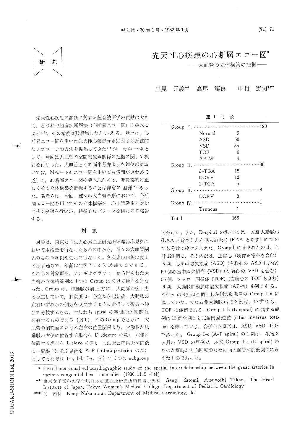
先天性心疾患の診断に対する超音波医学の貢献は大きく,とりわけ超音波断層法(心断層エコー図)の導入により1,2),その精度は数段増したといえる。我々は,心断層エコー図を用いた先天性心疾患診断に対する系統的なアプローチの方法を提唱してきた3,4)が,その一環として,今回は大血管の空間的位置関係の把握に関して検討を行なった。大血管とくに両半月弁よりも遠位部においては,Mモード心エコー図を用いても情報がきわめて乏しく,心断層エコー図の導入以前には,非侵襲的に正しくその立体構築を把握することは非常に困難であった。著者らは,今回,種々の大血管奇形において,心断層エコー図を用いてその立体構築を,心血管造影と対比させて検討を行ない,特徴的なパターンを得たので報告する。
The spatial structure between the great arteries (GA's) was studied in 165 cases with various congenital heart diseases (CHD) by using two-dimensional echocardiography (2-DE) com-pared with angiocardiography. All cases were divided into 4 groups according to the spatial structure from the angiocardiographic findings. Group I : anterior pulmonary artery (PA) and posterior aorta (Ao) as spiral interrelations. I-a ; right sided Ao and left sided PA as D-spiral.I-b ; left sided Ao and right sided PA as L-spiral. I-c ; Ao is just posterior to PA as AP-spiral. Group II : anterior Ao and posterior PA as paral-lel interrelations. II-a ; right sided Ao and left sided PA as D-parallel. II-b ; left sided Ao and right sided PA as L-parallel. II-c ; Ao is just anterior to PA as AP-parallel. Group III: Ao and PA are positioned as side by side (S. by S.).III-a ; right sided Ao and left sided PA as D-S by S. III-b; left sided Ao and right sided PA as L-S by S. Group IV : miscellaneous.
The ultrasonic planes used for this study are short axis views based on the position of the posterior GA named+1 to +4, i. e, plane+1 is the plane where the posterior semilunar valve is well visualized, +2 is slightly cephalic plane from +1 where the anterior semilunar valve is well seen, +3 is the plane where the posterior extension of one of the GA's is recognized initially, and +4 is the plane where the other GA is also extended posteriorly. The of which echo is initially extended can be identified as PA andthe other one is the Ao as we documented before. The 2-DE findings of each group are as following (Table 3). Group I. I-a ; two semi-lunar valves are seen at right postero-inferior and left antero-superior at the level of +1 and +2 respectivelly. Left anterior GA is extended posteriorly at +3 and right posterior GA is also extended at +4 toward left posterior or right posterior according to side of the aortic arch. I-b ; just mirror image to I-a. I-c ; two semilunar valves are seen postero-inferior and antero-superiorly at +1 to +2 respectively, and posterior extension of the anterior GA is see at +3. Group II : II-a ; two semilunar valves are seen at left postero-inferior and right antero-superior at +1 to +2 respectively. Posterior extension of the posterior GA is seen at +3. II-b ; just mirror image to II-a. II-c ; two semilunar valves are seen in postero-inferior and antero-superior directly at +1 to +2 respectively, and posterior extension of the posterior GA is visualized at +3. Group III: two semilunar valves are seen at the same level and the same depth at plane +2. III-a ; left sided GA is extended posteriorly at +3. III-b ; right sided GA is extended posteriorly at +3. Group IV : The 2-DE findings of Truncus arteriosus communis (type I) are shown as the miscellaneous group. Only a large GA is seen at +1, the distance between anterior and posterior wall becomes shortened at the central portion of the GA at +2, diverging into two GA's at +3 and left sided GA is extended left posteriorly at +4. Thus, piling up the echo images from +1 to +4 and reconstructing the three dimensional structural image, our method is very useful for diagnosis of CHD.

Copyright © 1982, Igaku-Shoin Ltd. All rights reserved.


