Japanese
English
- 有料閲覧
- Abstract 文献概要
- 1ページ目 Look Inside
- 参考文献 Reference
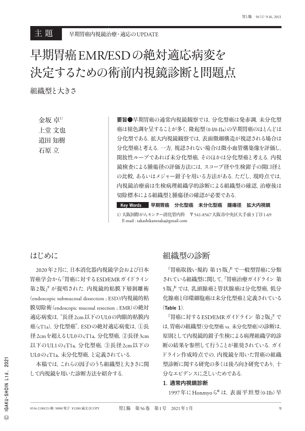
要旨●早期胃癌の通常内視鏡観察では,分化型癌は発赤調,未分化型癌は褪色調を呈することが多く,隆起型(0-I/0-IIa)の早期胃癌のほとんどは分化型である.拡大内視鏡観察では,表面微細構造が視認される場合は分化型癌と考える.一方,視認されない場合は微小血管構築像を評価し,開放性ループであれば未分化型癌,そのほかは分化型癌と考える.内視鏡検査による腫瘍径の評価方法には,スコープ径や生検鉗子の開口径との比較,あるいはメジャー鉗子を用いる方法がある.ただし,現時点では,内視鏡治療前は生検病理組織学的診断による組織型の確認,治療後は切除標本による組織型と腫瘍径の確認が必要である.
In white-light endoscopy for diagnosing early gastric cancer, differentiated-type cancer often appears reddish in color, whereas undifferentiated-type cancer appears pale. Elevated-type(0-I/0-IIa)cancer is typically a differentiated-type cancer. Furthermore, in magnifying endoscopy for diagnosing early gastric cancer, the combination of an absent microsurface pattern and opened-loop type microvessels is a feature of undifferentiated-type cancer, whereas a present microsurface pattern and/or polygonal/closed-loop microvessels are features of differentiated-type cancer. To estimate the tumor size using endoscopy, comparing the scope diameter and opening diameter of biopsy forceps, or using measure forceps, is recommended. Additionally, it is necessary to confirm the histological subtype by biopsy before endoscopic treatment and to confirm the tumor size by histological examinations using a resected specimen after endoscopic treatment.

Copyright © 2021, Igaku-Shoin Ltd. All rights reserved.


