Japanese
English
- 有料閲覧
- Abstract 文献概要
- 1ページ目 Look Inside
- 参考文献 Reference
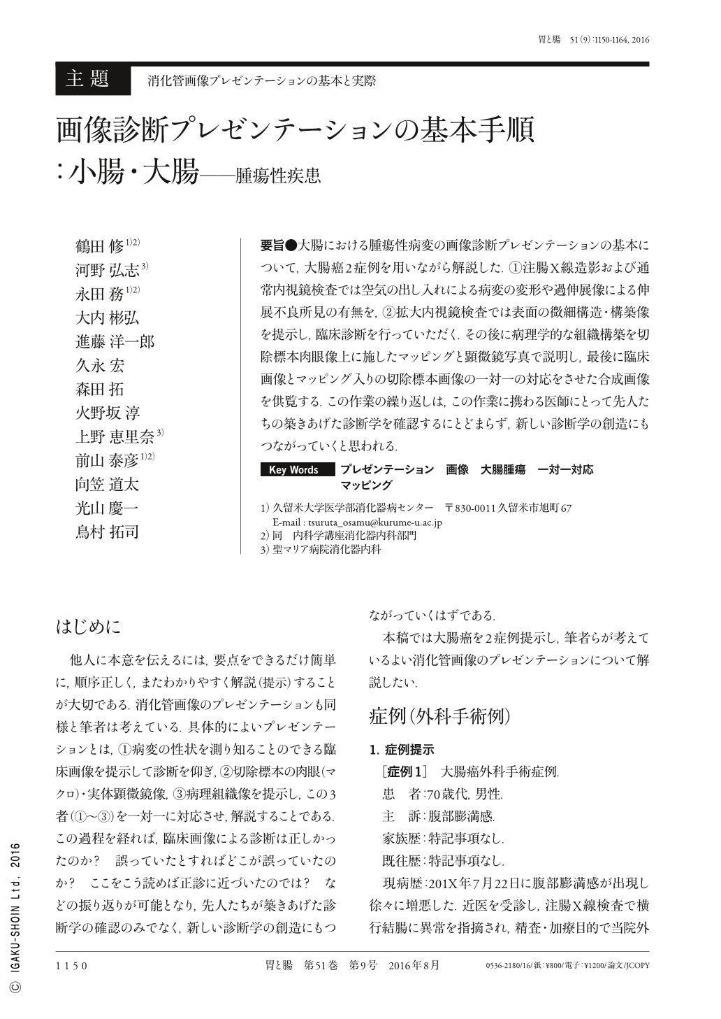
要旨●大腸における腫瘍性病変の画像診断プレゼンテーションの基本について,大腸癌2症例を用いながら解説した.①注腸X線造影および通常内視鏡検査では空気の出し入れによる病変の変形や過伸展像による伸展不良所見の有無を,②拡大内視鏡検査では表面の微細構造・構築像を提示し,臨床診断を行っていただく.その後に病理学的な組織構築を切除標本肉眼像上に施したマッピングと顕微鏡写真で説明し,最後に臨床画像とマッピング入りの切除標本画像の一対一の対応をさせた合成画像を供覧する.この作業の繰り返しは,この作業に携わる医師にとって先人たちの築きあげた診断学を確認するにとどまらず,新しい診断学の創造にもつながっていくと思われる.
Two cases of colorectal cancer were used to describe the basics of diagnostic imaging presentations of neoplastic lesions of the large bowel. Clinical diagnoses involve(1)assessing the bowel for poor elasticity resulting from changes in the lesion or hyperextension when air is introduced and removed during a barium enema examination or normal endoscopy examination and(2)examining the microstructures and other surface structures using magnification endoscopy. The pathological tissue structure is then described based on mapping the macroscopic images of the resected specimens and photomicrographs. Finally, composite images comparing(one-to-one)clinical images and images with mapping of the resected specimens are then obtained. Performing these procedures repeatedly will not only allow doctors involved to become highly familiar with the diagnostic process established by our predecessors but could also open up new avenues for the diagnostic techniques.

Copyright © 2016, Igaku-Shoin Ltd. All rights reserved.


