Japanese
English
- 有料閲覧
- Abstract 文献概要
- 1ページ目 Look Inside
- 参考文献 Reference
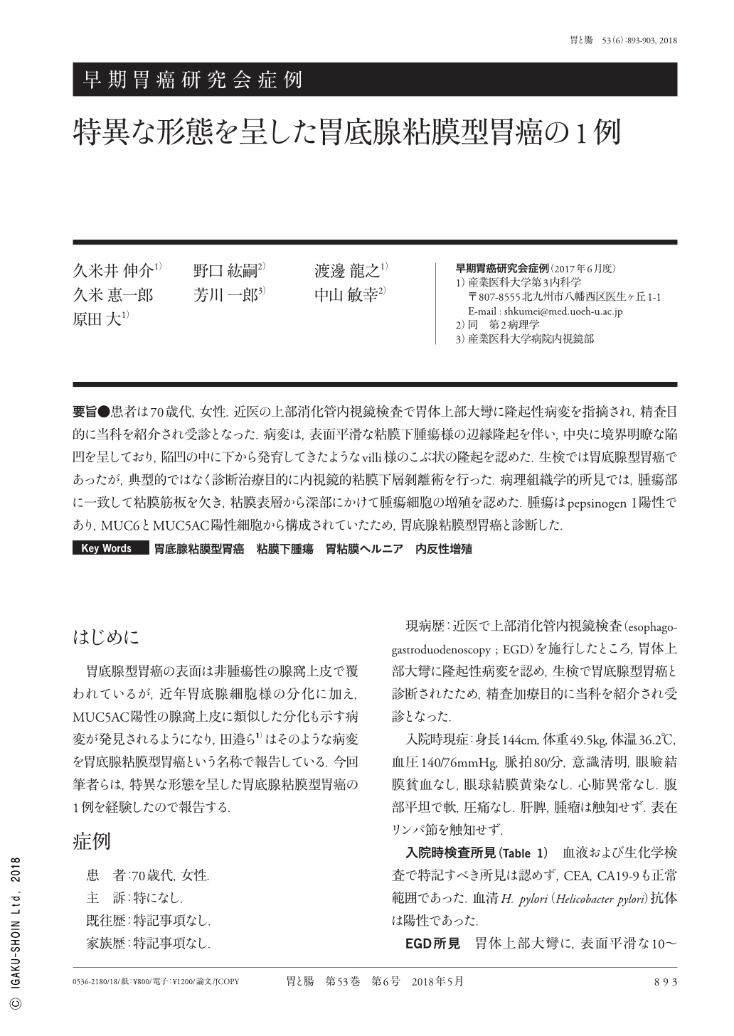
要旨●患者は70歳代,女性.近医の上部消化管内視鏡検査で胃体上部大彎に隆起性病変を指摘され,精査目的に当科を紹介され受診となった.病変は,表面平滑な粘膜下腫瘍様の辺縁隆起を伴い,中央に境界明瞭な陥凹を呈しており,陥凹の中に下から発育してきたようなvilli様のこぶ状の隆起を認めた.生検では胃底腺型胃癌であったが,典型的ではなく診断治療目的に内視鏡的粘膜下層剝離術を行った.病理組織学的所見では,腫瘍部に一致して粘膜筋板を欠き,粘膜表層から深部にかけて腫瘍細胞の増殖を認めた.腫瘍はpepsinogen I陽性であり,MUC6とMUC5AC陽性細胞から構成されていたため,胃底腺粘膜型胃癌と診断した.
A 70-year-old woman was referred to our hospital for undergoing further evaluation of a gastric lesion. The lesion was located at the greater curvature of the gastric body ; it was an elevated lesion and had a subepithelial tumor like-appearance and a clear depression at the top. A villi-like elevated tumor was noted in the depression area. Although a histological examination of the biopsy sample revealed a gastric adenocarcinoma of the fundic gland type, the lesion had atypical endoscopic findings. We performed endoscopic submucosal dissection as a therapeutic diagnosis. Histologically, adenocarcinoma of the fundic gland type without lamina muscularis mucosae was observed. Immunohistochemical analysis revealed that the tumor cells stained positive for Pepsinogen I, MUC6, and MUC5AC. Thus, this lesion was diagnosed as a gastric adenocarcinoma of the fundic mucosa type.

Copyright © 2018, Igaku-Shoin Ltd. All rights reserved.


