Japanese
English
- 有料閲覧
- Abstract 文献概要
- 1ページ目 Look Inside
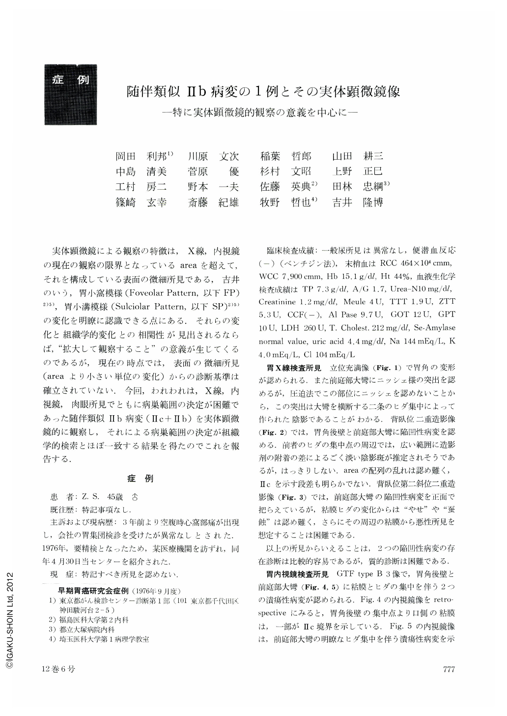
実体顕微鏡による観察の特徴は,X線,内視鏡の現在の観察の限界となっているareaを超えて,それを構成している表面の微細所見である,吉井のいう,胃小窩模様(Foveolar Pattern,以下FP)2)5),胃小溝模様(Sulciolar Pattern,以下SP)2)5)の変化を明瞭に認識できる点にある.それらの変化と組織学的変化との相関性が見出されるならば,“拡大して観察すること”の意義が生じてくるのであるが,現在の時点では,表面の微細所見(areaより小さい単位の変化)からの診断基準は確立されていない.今回,われわれは,X線,内視鏡,肉眼所見でともに病巣範囲の決定が困難であった随伴類似Ⅱb病変(Ⅱc+Ⅱb)を実体顕微鏡的に観察し,それによる病巣範囲の決定が組織学的検索とほぼ一致する結果を得たのでこれを報告する.
One “area gastrica” comprises a number of Foveolar Pattern (abbreviated to FP below) and Sulciolar Pattern (abbreviated to SP below), which were first advocated by Prof. Yoshii. They could play a great part in the estimation of existence and nature of lesion.
So far, attentions have been mainly paid to alterations in the level of “area gastrica” radiographically and endoscopically. Stereomicroscopic observation, however, is characterized by the fact that beyond “area gastrica” alterations of more minute units (FP and SP) are clearly recognizable. They correspond to gastric pits on a section.
We observed a case of concomitant IIb-like lesion of the stomach stereomicroscopically, of which precise extension was difficult to determine radiographically, endoscopically and macroscopically. The extent of the lesion confirmed by stereomicroscope corresponded well with the result of histologic observation. IIb-like lesion was well demarcated from the surrounding normal mucosa revealing well maintained SP. Compared with normal SP, appearance of the IIb-like lesion was rather monotonous, caused by carcinomatous invasion which resulted in destruction and consequent decrease of SP. Destroyed appearance on a surface might have some connection with the type of carcinoma cell (predominant mucocellular adenocarcinoma in the present case).
Blurring and disappearance of SP were here and there recognizable in the lesion. Their degree could be well accounted for by the section involved. Sections around IIb-like lesion showed three different types of carcinomatous invasion; carcinoma is exposed to the surface itself; carcinoma is covered with only one layer of regenerative epithelium; or carcinoma is confined within the lamina propria maintaining its basic structure of the mucous membrane.
Now, we would like to mention below a few affiliated problems with Stereomicroscopic observation. First of all, correlation between severity of blurring and disappearance as superficial alterations and its histological changes as a background should be called in question, especially in case carcinomatous invasion is confined only within the lamina propria with a basic structure of the mucous membrane well maintained.
Secondly, there exists a certain probability that FP and SP could be effective not only in quantitative diagnosis but also in qualitative diagnosis of a lesion. In other words, superficial alterations of FP and SP in differentiated carcinoma are supposed to be different from those in poorly differentiated carcinoma. On a surface of differentiated carcinoma, minute ditch-like structure with irregularity resulting from malignant gastric pit, villoid unevenness and comparatively plain FP or SP similar to a crab hole are to be recognized. Although the structure is preserved, the surface of a differentiated carcinomatous lesion tends to reveal extremely desolate appearance, while that of a poorly differentiated carcinomatous lesion is apt to show blurring and disappearance of FP and SP, giving an impression of destruction.
Thirdly, stereomicroscopic systematization with alterations of FP and SP in what it terms “chronic gastritis” is also to come into our mind, related to advent of carcinoma.
Finally, we should impart a sufficient meaning to superficial “observation by magnification” prior to the improvement of dye-combined endoscopy and magnifying instrument. Summing up, we are now in an initial stage of finding out a significance in “observation beyond area gastrica”.

Copyright © 1977, Igaku-Shoin Ltd. All rights reserved.


