Japanese
English
- 有料閲覧
- Abstract 文献概要
- 1ページ目 Look Inside
要旨 微小胃癌の診断限界を知るため切除胃を実体顕微鏡で観察後,病理組織学的検索をした633例について,大きさを(A)3mm未満,(B)3≦~≦5mm,(C)5<~≦10mm,に分け発見方法を臨床検査,実体顕微鏡観察および病理組織学的検索とに分類し検討した.臨床,実体までを診断可能とすると,(B)は29病変中17病変,発見率59%,(A)は19病変中2病変11%と低い.表面の形態を実体顕微鏡でみると5,4mmでははっきりした癌の特徴をとるが,3mmではわずかになり,2mmでははっきりしない.以上より3mmの大きさが診断限界と考えた.領域別では幽門腺領域で40病変中18病変(45%),胃底腺領域で8病変中1病変(13%)と発見率は低い.胃底腺領域の微小胃癌の発見はかなり困難である.
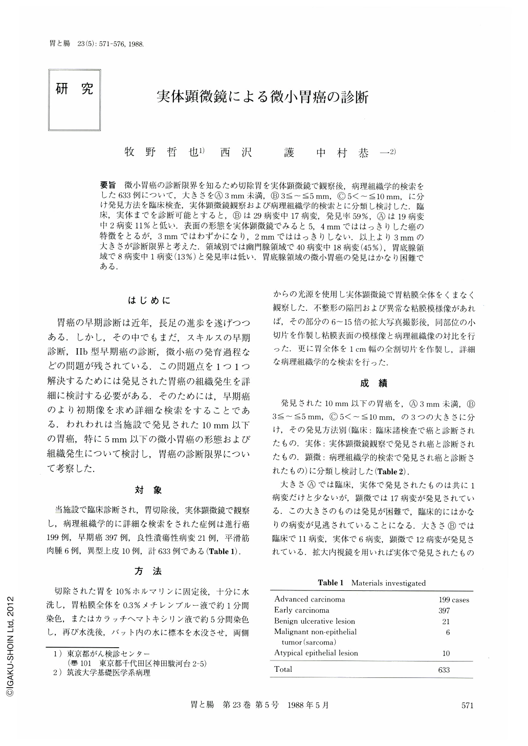
In order to define the diagnostic limit of microcarcinoma, stereomicroscopic observation of the resected parts of the stomach was performed by using dyestaining method in 596 cases with gastric carcinoma, 21 cases with benign ulcerative lesion, 6 cases with malignant non-epithelial tumor and 10 cases with atypical epithelial lesion. This was carried out prior to histopathological examination.
12 microcarcinomas less than 5 mm in diameter and 27 minute carcinomas between 5 and 10 mm in diameter were detected before surgery. Using stereomicroscopy, a further 7 microcarcinomas and 2 minute carcinomas were able to be located. Another 29 microcarcinomas and 20 minute carcinomas were found by histological examination.
If the microcarcinomas detected by the stereomicroscopy could have been detected by using magnifying endoscopy, 58.6% of a total of 29 microcarcinomas between 3 and 5 mm in diameter and 59.2% of a total of 49 minute carcinomas between 5 and 10 mm in diameter would have been clinically detectable.
Detectability, however, of microcarcinomas less than 3 mm in diameter was only 11% of the total, and these foci were hardly recognizable even when using a magnifying instrument.
Stereomicroscopic features of malignancy could be identified in the microcarcinomas more than 3 mm in diameter, but only a few of them could be noted in the foci 3 mm in diameter. In the foci less than 3 mm in diameter, discrimination between malignancy and benignancy was extremely difficult.
Thus, only about 60% of microcarcinomas more than 3 mm in diameter, and 60% of minute carcinomas can possibly be detected by using a magnifying endoscope. It can also be said that the possibility of diagnosing microcarcinoma of the stomach is limited to those carcinomas not below 3 mm in diameter.
Microcarcinomas in the pyloric gland area were rather easily detected with the detectability of 45% but difficulty arises in the detection of microcarcinomas in the fundic gland area with a detectability of 13%.
Copyright © 1988, Igaku-Shoin Ltd. All rights reserved.