Japanese
English
- 有料閲覧
- Abstract 文献概要
- 1ページ目 Look Inside
- サイト内被引用 Cited by
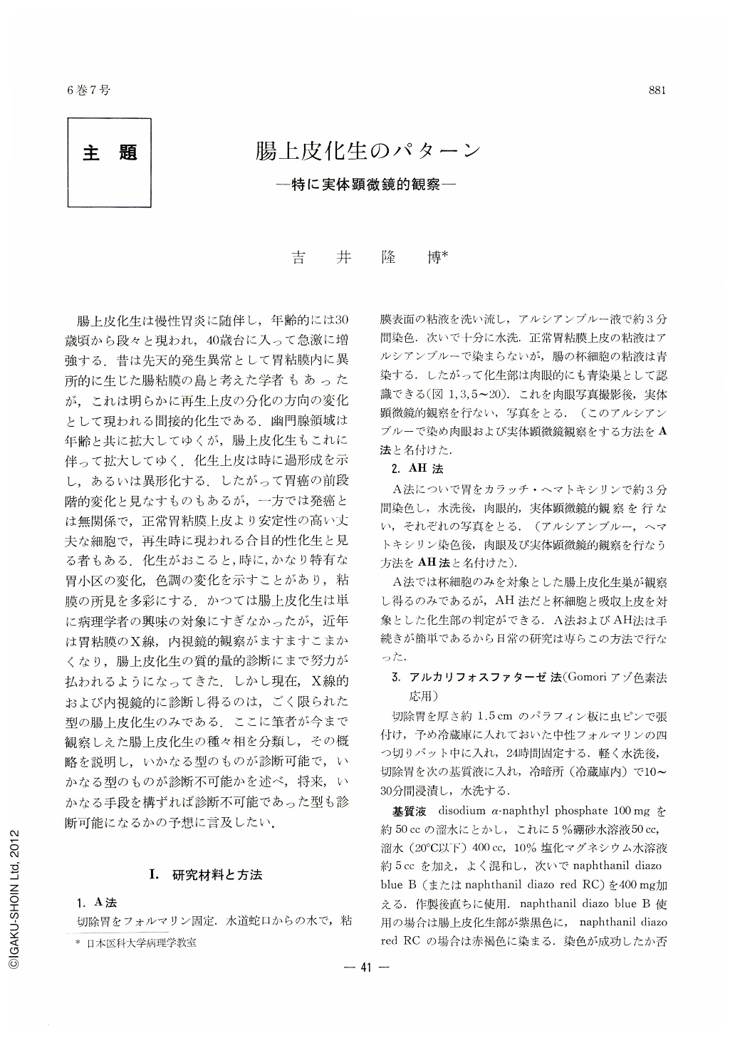
腸上皮化生は慢性胃炎に随伴し,年齢的には30歳頃から段々と現われ,40歳台に入って急激に増強する.昔は先天的発生異常として胃粘膜内に異所的に生じた腸粘膜の島と考えた学者もあったが,これは明らかに再生上皮の分化の方向の変化として現われる間接的化生である.幽門腺領域は年齢と共に拡大してゆくが,腸上皮化生もこれに伴って拡大してゆく.化生上皮は時に過形成を示し,あるいは異形化する.したがって胃癌の前段階的変化と見なすものもあるが,一方では発癌とは無関係で,正常胃粘膜上皮より安定性の高い丈夫な細胞で,再生時に現われる合目的性化生と見る者もある.化生がおこると,時に,かなり特有な胃小区の変化,色調の変化を示すことがあり,粘膜の所見を多彩にする.かつては腸上皮化生は単に病理学者の興味の対象にすぎなかったが,近年は胃粘膜のX線,内視鏡的観察がますますこまかくなり,腸上皮化生の質的量的診断にまで努力が払われるようになってきた.しかし現在,X線的および内視鏡的に診断し得るのは,ごく限られた型の腸上皮化生のみである.ここに筆者が今まで観察しえた腸上皮化生の種々相を分類し,その概略を説明し,いかなる型のものが診断可能で,いかなる型のものが診断不可能かを述べ,将来,いかなる手段を構ずれば診断不可能であった型も診断可能になるかの予想に言及したい.
Ⅰ. After resected stomachs were fixed with formalin and stained with alcian blue (A method) and alcian blue-hematoxylin (AH method), intestinal metaplasia was investigated macroscopically, dissecting-microscopically and histologically and was classified as follows. : From macroscopic distribution;①discrete type, ②specific type (Takemoto type), ③erosion type, ④linear and ramified type, ⑤geographic type and ⑥diffuse type. From relationship to areae gastricae; ①area-unit type, ②sulcus type (intestinal metaplasia in marginal furrows of areae gastricae) and ③irregular type (distribution without relationship to areae gastricae or furrows).
Ⅱ. Dissecting-microscopically, metaplastic mucosa, as a rule, shows sulciolar pattern (named by author, see Fig. 26), which is coarser than of non-metaplastic mucosa (Fig. 21, 22) and which frequently demonstrates mesh-like pattern.
Ⅲ. The author classified areae gastricae as follows.: 1) round (R), 2) irregular (I) and 3) sausagelike (S) areae. Each type was subdivided by size into three subgroups.; 1) large (1), 2) medium-sized (m) and small (s). Each type was also subdivided by grade of protrusion into three; 1) markedly protruded (P2), 2) slightly protruded (P1) and 3) fiat (f). Areae gastricae completely encircled by marginal furrows are marked with “Comp” and those incompletely encircled are marked with “Incomp”. Intestinal metaplasia was found in all kinds of areae gastricae, so that metaplasia could not be diagnosed grossly from the form of areae gastricae with one exception. That is the specific type which can be diagnosed radiologically, endoscopically and macroscopically. This type has four characteristics. Namely, areae gastricae with specific type of metaplasia are 1) conspicuously larger, 2) more protruded and 3) more grayish-white than non-metaplastic areae and 4) disseminated as discrete type. If these four characteristics are well understood, intestinal metaplasia can more accurately be diagnosed even when they have become less distinct. With decreasing characteristics, the diagnosis naturally becomes less accurate.
Ⅳ. If these methods (A and AH) and the abovedescribed findings could be utilized in endoscopic examination, especially in high-power fibergastroscopic study now being developed, it would become possible to diagnose not only the specific type but also any other types of intestinal metaplasia.

Copyright © 1971, Igaku-Shoin Ltd. All rights reserved.


