Japanese
English
- 有料閲覧
- Abstract 文献概要
- 1ページ目 Look Inside
隆起性病変のX線診断は,その形態から,大きさ,高さ,側面像,正面像をもって,Ⅱa,ATP,ポリープ,粘膜下腫瘍の鑑別診断を行なっている.その診断結果を,西沢は,X線検査で悪性だと診断し,組織も悪性であったものが82.1%,良性と診断し,組織も良性であったものが97.3%と報告した.X線検査と内視鏡検査を行ない,その最終診断でも悪性の疑いとして残ったものが7.9%,また,良性だと誤診したものが6.3%と,われわれも報告した.ここに生検を行なわなくてはならない領域がある.
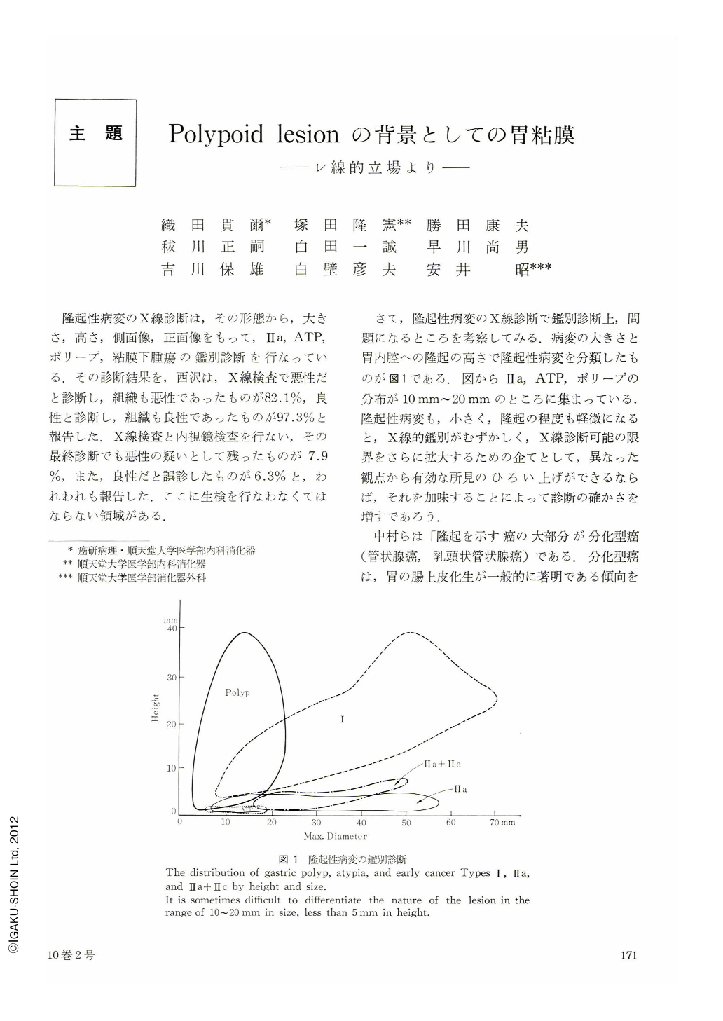
さて,隆起性病変のX線診断で鑑別診断上,問題になるところを考察してみる.病変の大きさと胃内腔への隆起の高さで隆起性病変を分類したものが図1である.図からⅡa,ATP,ポリープの分布が10mm~20mmのところに集まっている.隆起性病変も,小さく,隆起の程度も軽微になると,X線的鑑別がむずかしく,X線診断可能の限界をさらに拡大するための企てとして,異なった観点から有効な所見のひろい上げができるならば,それを加味することによって診断の確かさを増すであろう.
X-ray examination differentiates the nature of a protruded lesion by its shape, size, and face-on and lateral views.
The smaller and lower the size and the height of the lesion become, the more difficult the differentiation with x-ray becomes, especially in the range of 10~20 mm in size.
If we pay our attention to the appearance of the surrounding mucosa of the lesion, it may become easier to differentiate the nature of a protruded lesion, and the x-ray appearances of the surrounding mucosa have been analysed.
This study led the author to the following conclusion:
1) X-ray appearance of the surrounding mucosa of an early cancer Type Ⅱa within the about double length of the greatest radius from the center of the protruded lesion shows marked granurality in the most cases.
And its marked intestinal metaplasies is histologically proved.
2) X-ray appearances of the surrounding mucosa of an atypia are less granuler than that of Type Ⅱa case.
In some cases, no intestinal metaplasia of the surrounding gastric mucosa was histologically defined.
3) In most cases with hyperplastic polyp, the surrounding mucosa is radiologically smooth and proved histologically to be mucosa with on evidence of intestinal metaplasia.
4) It is double contrast study that we can delineate the each walls of the stomach, then to delineate the appearance of the surrounding mucosa radiologically, double contrast method is better than compression study.

Copyright © 1975, Igaku-Shoin Ltd. All rights reserved.


