Japanese
English
- 有料閲覧
- Abstract 文献概要
- 1ページ目 Look Inside
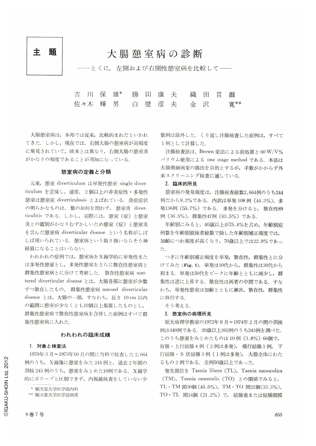
大腸憩室病は,本邦では従来,比較的まれだといわれてきた.しかし,現在では,右側大腸の憩室病が高頻度に発見されていて,欧米とは異なり,右側大腸の憩室炎がかなりの頻度であることが周知になっている.
憩室病の定義と分類
元来,憩室diverticulumは単発性憩室single diverticulumを意味し,通常,2個以上の非炎症性・多発性憩室は憩室症diverticulosisとよばれている.炎症症状の明らかなものは,数の如何を問わず,憩室炎diverticulitisである.しかし,実際には,憩室(症)と憩室炎との鑑別がかなりむずかしいため憩室(症)と憩室炎を含んだ憩室病diverticular diseaseという名称がしばしば用いられている.憩室病という取り扱いならそう神経質になることはいらない.
Diverticular disease of the colon had been a rare condition in Japan. But, with the improvement of x-ray examination and increase in the number of patients examined its frequency became exceedingly high in recent years.
The authors had 244 cases with diverticular disease of the colon from March, 1970 through October, 1973. The incidence was 9.2% out of the examined 2,664 cases. These 244 cases included 108 cases (44.3%) with single diverticular disease, 89 cases (36.5%) with scattered diverticular disease and 47 cases (19.2 %) with massed diverticular disease.
Age: Single diverticular diseases were observed in patients over 10, but massed diverticular diseases were observed in patients over 30 and 52.3% of massed diverticular diseases were observed in patients over 60. That is, single diverticular disease will gradually develope to scattered diverticular disease and massed diverticular disease. The average age of the rightsided massed diverticular disease was 54.3, while it was 66.4 in the left-sided massed diverticular disease. Location : About 70% of the cases with single diverticular diseases were localized at the right colon (ascending coin 43.0%, coecum 30.8%) and a few at the left colon (sigmoid colon 4.7%, descending colon 9.2%) except transverse colon. Massed diverticular diseases were observed in the right colon in 68.1% of the cases, the left colon in 29.8% and entire colon in 2.1%, The high incidence of diverticular disease of the right colon among the Japanese suggests that there is a hereditary or racial predisposition to the disease.
Pathological findings : In the 243 cases of autopsy, there were 10 cases with diverticular disease (66 diverticula), 4 cases were localized at the coecum and ascending colon, 3 cases at the sigmoid colon and descending colon, 1 case at the transverse colon, and 2 cases at the entire colon. Diverticular were observed in 9.1% (6 out of 66 diverticula), and almost of them (5 out 6 diverticula) were, recognized at the right-sided diverticular disease with over 10 diverticular. The thickness of circular muscle was 2.2~2.5 mm in sigmoid diverticular disease, and 0.9~1.0 mm in coecum and ascending colon diverticular disease. Deformity of diverticulum was related to the grade of inflammatory change.
X-ray findings : Cases with right-sided massed diverticular, disease showed marginal irregularity in 36.7 %, haustral irregularity in 43.3%, limited distensibility in 56.7%, crowded folds in 43.3% and asymmetrical folds in 6.7%. On the other hand, cases with massed diverticular disease located at the left colon showed marginal irregularity in 78.6%, haustral irregularity in 85.7%, limited distensibility in 78.6%, crowded folds in 85.7% and asymmetrical folds in 57.1%. The difference of x-ray findings between right-sided and left-sided massed diverticular drisease could be caused by the difference of muscular thickening.
We compared the differences between the right sided and the left sided on x-ray findings of 73 diverticulitis (12 of the right colon and 61 of the left colon) reported in literatures. The following data are shown about (a) the right colon and (b) the left colon. Marginal irregularity: (a) 58.3%, (b) 65.6%, deformity: (a) 33.3% (b) 85.2%, limited distensibility: (a) 66.7% (b) 82.0%, crowded folds (a) 8.3% (b) 29.5%, irregular folds: (a) 41.7% (b) 54.0%, cushion-like defect : (a) 58.3% (b) 52.5 %, fistula: (a) 0% (b) 57.4%, abscess-cabity: (a) 0% (b) 39.3%and barium fleck: (a) 8.3% (b) 19.7%.
Mild findings are seen in the right colon comparing in the left. It is reported that it is based on anatomical and pathological differences.

Copyright © 1974, Igaku-Shoin Ltd. All rights reserved.


