Japanese
English
- 有料閲覧
- Abstract 文献概要
- 1ページ目 Look Inside
腺癌の頸部リンパ腺転移を認めた患者の原発巣を検索し,胃体部にⅡc様の所見を認め,そこからの胃生検で癌を診断し,剖検にてこれが原発巣であったことを確認した症例を経験したので報告する.
Cervical lymph node metastasis of adenocarcinoma variety in a patient recently examined led us to the discovery of a Ⅱc-like lesion in the gastric corpus. Biopsy demonstrated cancer within it, later confirmed at autopsy as the original site of cancer. However, as both roentgenogram and endoscopy revealed nothing but early-cancer-like finding there, it was hardly possible to determine this lesion as the primary site of cancer.
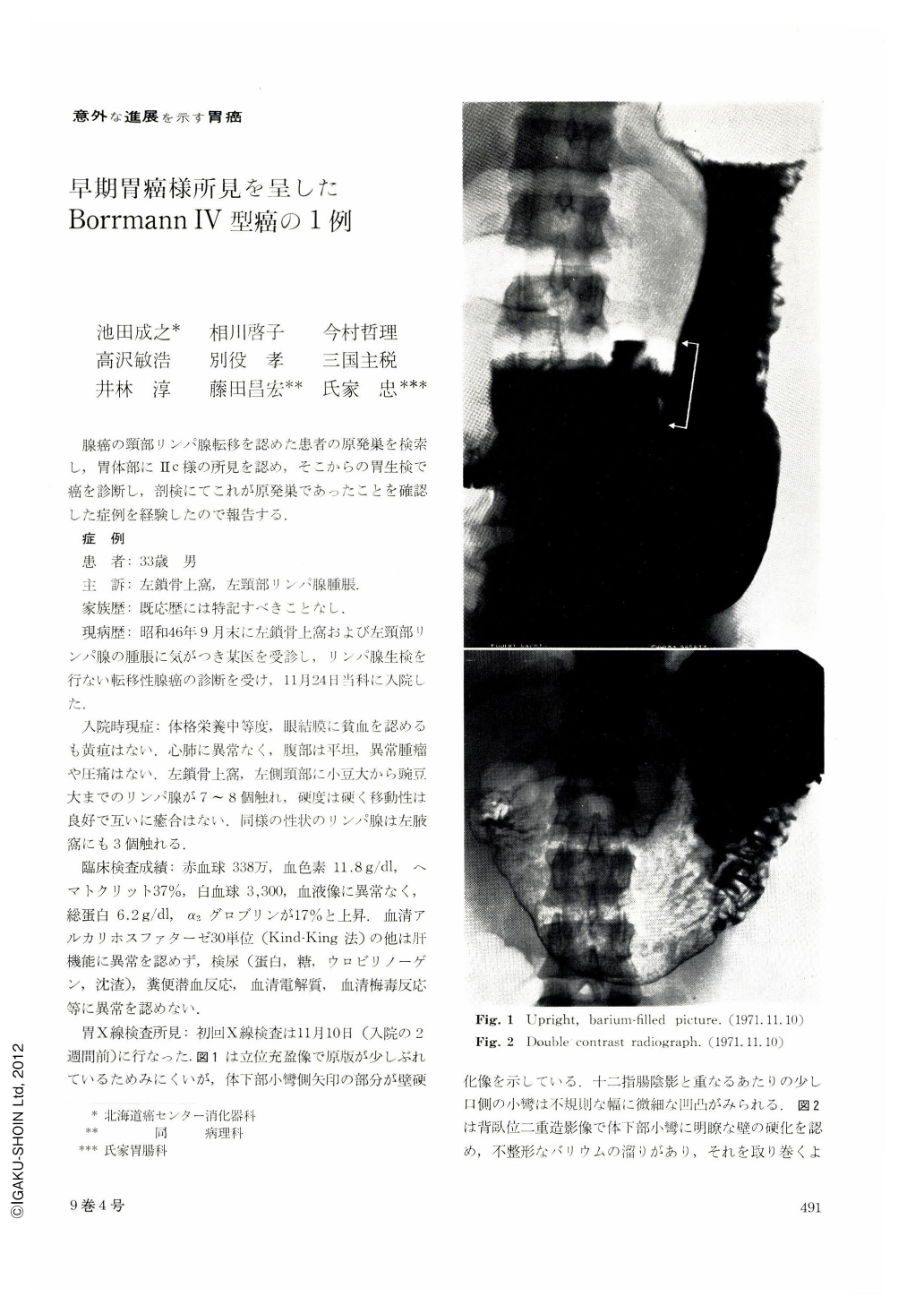
The patient, a 33-year-old man, came to us with a chief complaint of swollen lymph nodes in the left supraclavicular region and left-hand side of the neck. Biopsy showed that the swelling was due to metastatic adenocarcinoma. The primary site of cancer was naturally looked for in the digestive tract including the stomach, intestine and gallbladder. In upright barium-filled picture of the stomach was found rigidity and irregularity of the wall in the lower part of the body. Mural rigidity was also noticed in double contrast picture on the lesser curvature of the same site. A barium pool of irregular shape was also visualized, surrounded with rough, granular mucosa. A diagnosis of Ⅱc was thus made. Gastric roentgenogram one month later showed that distensibility of the stomach as a whole was hardly altered; and in double contrast picture there was too much of mucus for the contrast medium to adhere to the mucosal surface. Nonetheless, swollen and tortuous mucosal folds on the posterior wall and the greater curvature side were such that we arrived at a diagnosis of Borrmann Ⅳ type cancer of the stomuch. Pictures of GTF-A examination done about the same time as the initial roentgenography showed several, reddened and granular spots on the lesser curvature of the lower body near the anterior wall. The mucosal folds converging toward the reddened area tapered off at their tips. Endoscopic diagnosis then was still Ⅱc. The resected stomach showed scar-like changes in the corpus along with general swelling of the rugae over the entire stomach, Histologically, the lesion belonged to adenocarcinoma tubulare mucocellulare. Only a small part of it was exposed on the mucosal surface, and most of it was seen as diffuse infiltration with interstitial hyperplasia in the submucosa, muscular coat and subserosa.
Copyright © 1974, Igaku-Shoin Ltd. All rights reserved.