Japanese
English
- 有料閲覧
- Abstract 文献概要
- 1ページ目 Look Inside
はじめに
胃内視鏡の進歩に伴い,これまで稀にしか経験することができず,しかも術前診断が非常に困難であった胃病変の発見も可能となり,その臨床診断についての検討もより詳細に行ないうるようになった.最近,筆者らは胃粘膜下囊胞とⅠ型早期胃癌が共存していたきわめて稀な症例を経験したので報告する.
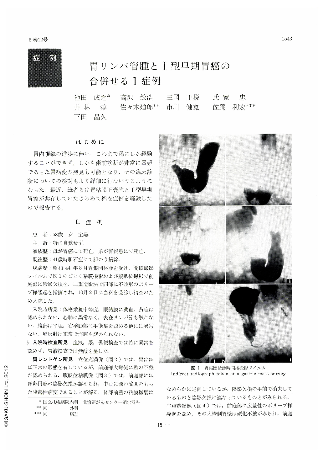
A housewife 58 years of age was referred for thorough examination of the stomach because of a shadow defect in the antrurn found at a gastric mass survey. She had been free from any subjective symptoms. The stomach was almost of normal contour in the barium-filled picture, but in the prone mucosal study an almost oval shadow defect was visualized in the antrum. In the double contrast picture it was seen as a broad-based polypoid elevation, with its surface rough and uneven. A deep depression was seen in its center. Besides this shadow defect, pressure films disclosed above the angle an oval rediolucency with relatively ill-defined margins. Gastric endoscopy revealed on the posterior wall of the antrum an irregular-surfaced polypoid elevation partly covered by white coat, with constriction at its base, together with a hemispheric protrusion on the anterior wall at the level of the angle. Of smooth surface, it was of the same color as the surrounding mucosa.
The polypoid lesion on the posterior wall of the antrum was diagnosed as adenocarcinoma papillare by gastric biopsy while the hemispheric elevation on the anterior wall of the angle was suspected as non-substantial submucosal tumor because at biopsy its surface was seen to yield easily to the tip of the forceps.
In the resected stomach a broad-based protrusion was recognized on the posterior wall 6cm oral from the pyloric ring, measuring 2×3×1.5 cm. Histologically it was papillar adenocarcinoma, a type Ⅰ early cancer partly infiltrating into the submucosal layer. On the anterior wall a little oral from this lesion was seen a hemispheric elevation, which measured 3×3×1 cm. It was a submucosal cystoma with uniform fluid accumulation in its cut sections. Histologically it was a monolocular cystoma with its wall consisting of a single layer of flat endothelial cells. It was considered as a retention cyst caused by obstruction and dilation of lymphatic vessels.
Copyright © 1971, Igaku-Shoin Ltd. All rights reserved.