Japanese
English
- 有料閲覧
- Abstract 文献概要
- 1ページ目 Look Inside
はじめに
早期胃癌の診断に関する研究は著しいものがあり,Ⅱb型早期胃癌についての報告も多数みられる時代になった.本症例は,切除胃肉眼所見上,病巣を指摘できるⅡb+Ⅱa型早期胃癌であるが,術前に確診しえたので報告する.
This is a case report of Ⅱb+Ⅱa type early gastric cancer, preoperatively diagnosed as such and later confirmed by gross observation of the resected specimen.
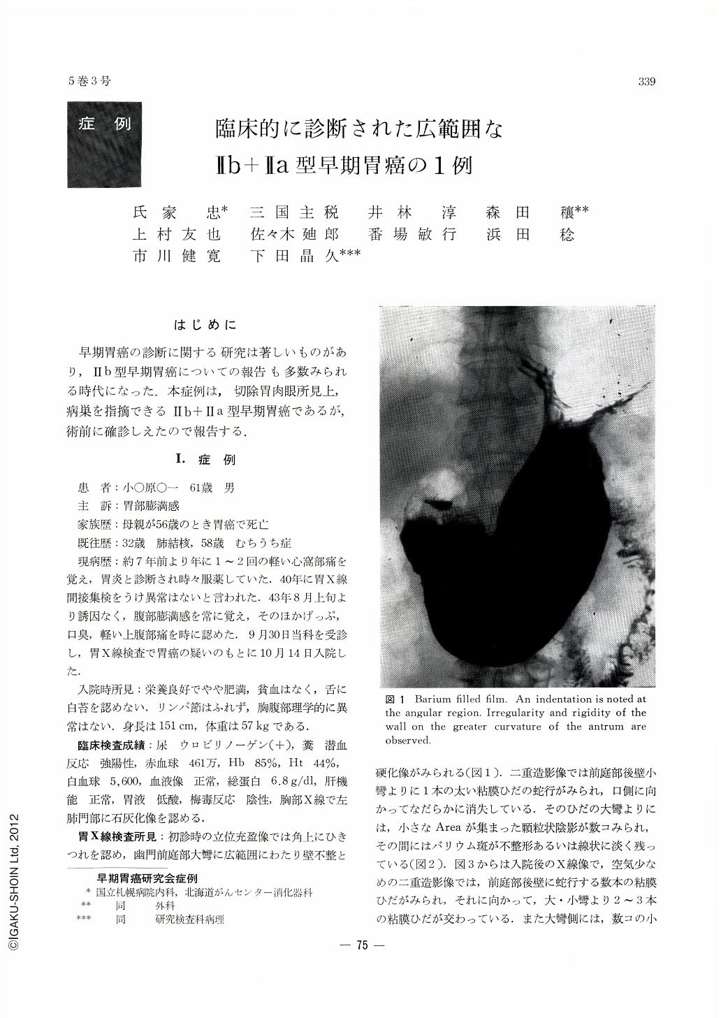
A 61-year-old man visited the authors' hospital on account of feeling of fullness in the epigastrium. As he was suspected by x-ray examination of harboring gastric cancer, he was admitted to the hospital. Laboratory data at the time of admission revealed no abnormality except strong positive occult blood in the feces. At x-ray study, extensive irregularity and rigidity of the gastric wall were noticed in upright, barium-filled films on the greater curvature of the pyloric antrum, while in double contrast study serpentine mucosal folds were visualized together with granular shadows consisting of irregular-sized areae gastrieae. In another double contrast study with lesser amount of air, several winding mucosal folds were demonstrated on the posterior wall of the antrum in addition to a few mucosal folds running crosswise to them. On compression, a comma-shaped radiolucency was visualized on the lesser curvature of the antrum. It was diagnosed as Ⅱb+Ⅱa. By endoscopic examination a small Ⅱa was recognized on the anterior wall of the antrum. By repeat study the antral mucosa was observed to be edematous and a slightly arched contraction was seen on the anterior wall and in the greater curvature side of the antrum. An area of discoloration was also recognized on the greater curvature with tiny, engorged spots scattered about within it. This finding was suspicious of Ⅱa+Ⅱb. The diagnosis of cancer was finally arrived at by taking tissue specimens chiefly from the Ⅱa lesion on the anterior wall of the antrum. Gross observation of the resected stomach showed that except a part of the posterior wall of the antrum the mucosal folds all around the antral lumen were serpentine with discoloration and loss of luster of the mucosal surface. The lesion measured about 7.5 by 4.0cm, extending from the anterior wall across the greater curvature way to the posterior wall. A Ⅱa was found in the lesion on the anterior wall of the antrum, measuring 1.3 by 0.8 cm. Macroscopically the lesion as a whole was classified as Ⅱa, but histopathologically no difference was found in mucosal thickness between this wide area of infiltration and the surrounding non-cancerous mucosal layers, so that it was diagnosed as Ⅱb+Ⅱa. It was adenocarcinoma tubulare with depth invasion limited within the mucosa. Even when retrospectively analyzed, the margins of the lesion could not be determined by endoscopy.
Some reference has been made to the diagnosis of Ⅱb type early gastric cancer.
Copyright © 1970, Igaku-Shoin Ltd. All rights reserved.